Download
1 / 27
270 likes | 634 Views
Knee Anatomy (1). Modified hinge joint flexion/ extension, internal/ external rotation Two distinct joints tibiofemoral joint Patellofemoral joint. Knee Anatomy (2). Tibiofemoral joint condyles of the femur very rounded medial condyle is larger than the lateral condyle Tibial plateaus

E N D
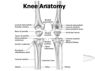
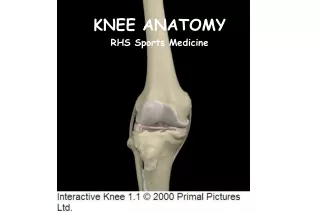
Knee Anatomy (1) • Modified hinge joint • flexion/ extension, internal/ external rotation • Two distinct joints • tibiofemoral joint • Patellofemoral joint
Knee Anatomy (2) • Tibiofemoral joint • condyles of the femur • very rounded • medial condyle is larger than the lateral condyle • Tibial plateaus • flattened, very slightly concave • “Screw home mechanism” • required to reach full extension • tibia rotates laterally on the femur to produce a locking of the knee
Knee Anatomy (3) • Patellofemoral joint • patella • triangular shaped seasamoid bone: protect the knee joint • femur • Patellofemoral groove or trochlear surface • Q angle • The angle of pull of quadriceps on the patella • normal is 13 degrees male/ 18 female
Knee Anatomy (4) • Menisci • firbrocartilage discs • Functions:1) deepen the tibial plateaus or joint2) absorption and dissipation of force3) congruency of the surface to improve wt distribution4) nourishment and lubrication of joint surfaces • Thicker along the lateral portion
Menisci Cont • Poor blood supply (only outer 1/3 receives direct blood supply) Fig 11-5-C • Medial is C shaped; Lateral is O shaped • The medial is more commonly injured because of its attachment to the MCL ligament & more securely attached to the tibia (which makes it less mobile)
Knee Anatomy (5) • 4 main ligaments- help stabilize knee jt • Medial Collateral (Tibial Collateral) • prevents valgus & rotational forces/stresses • Attaches to medial femoral epicondyle and anterior medial tibia • Lateral Collateral (Fibular Collateral) • prevents varus struss • Attaches to lateral femoral epicondyle and head of fibula
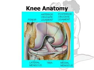
Knee Anatomy (6) Fig 11-9 • Anterior Cruciate (ACL) • Prevents tibia from moving forward/ femur from going back • attaches to lateral femoral condyle/ medial tibia at intercondylar eminence • Posterior Cruciate (PCL) • Prevents tibia from moving backward/ femur from going forward • attaches to medial femoral condyle/ lateral tibia at intercondylar eminence
Knee Anatomy (7) • Bursa – Fig 11-2 C • formed by joint capsule • function to reduce friction • several: • Suprapatellar: largest in body • Prepatellar: between skin and patellar tendon (housemaids knee) • Infrapatellar: below petella (superficial and deep) • Pes anserine bursa- medial proximal aspect of tibia
Knee Anatomy (8) • Muscles-contribute to jt stability • Quadriceps (EXT): Vastus lateralis, vastus medialis, rectus femoris, vastus intermedius; quads also aid in patella alignment • Hamstrings (Flex): Semitendinosus (IR), Semimembranosus(IR), Biceps Femoris (ER) • Gastroc (Flex), Sartorius(Flex/IR), Gracilis (Flex/IR), & popliteus (Flex)
Knee Anatomy (9) • Blood supply – Fig 11-5 • femoral artery to popliteal artery, then medial superior/inferior genicular, lateral superior/inferior genicular • Nerve Supply • Femoral nerve(Ant); Sciatic nerve (post) to tibial nerve and common peroneal nerve
Prevention of Knee Injuries • Stretching and strengthening of knee (FS 11.1) • Protective Knee BracesThree types: prophylactic, functional, and rehabilitative (Fig 11-6) • Patellofemoral- Fig 11-7- “Cho-Pat” strap: horseshoe knee sleeve • Proper footwear- correct shoe for the correct surface
Treatment of Knee Injuries • Normal acute protocol and NSAIDs • Progression of cold to hot treatments • Control swelling, fit for crutches if necessary,increase ROM and strength • Return to competition the safest and quickest way possible thru rehab, functional activities, and sports specific activities
MCL Injuries • MOI: valgus stress or lateral forces, internal rotation • HOPS • Pain and swelling over the medial joint, • pn over medial epicondyle or medial tibia, • + valgus stress test • Tx • hinged knee brace, treat symptoms, strengthen musculature, rule out meniscus tear with MRI; will heal by itself with conservative treatment; immobilize
LCL Injuries • MOI: Varus stress or medial forces • HOPS • Pain and swelling over the lateral joint, • pn over lateral epicondyle or fibular head, • + varus stress test • Tx • hinged knee brace, treat symptoms, strengthen musculature; immobilize; can heal by itself
ACL Injuries • MOI: • Sudden deceleration, blow to lateral leg with the knee bent, foot fixed • HOPS • Immediate pain and swelling; hot knee; Pain “inside the knee”; knee “feels loose”, “something not right” • + anterior drawer stress test and lachmans • Tx • depends on the severity, with 3rd degree = surgery; treat symptoms; immobilize
PCL Injuries • MOI: • Fall on a bent knee; posterior force on tibia, hyperextension • HOPS • Immediate pain and swelling; hot knee; Pain in the popliteal fossa; knee “falling apart” knee “feels loose” • + posterior drawer stress test, posterior sag test • Tx • depends on the severity, immobilized, strengthen knee musculature; surgery
Menisci Injuries • MOI: Twisting with foot fixed • HOPS • Pn over the joint line, catching/locking or giving out of the knee. Popping or clicking in joint line, swelling after activity with little heat, Pn with or deep squat • Tx • strengthen knee musculature, surgery if sx persist; recovery time depends on type of surgery and tear
Patello Femoral Stress Synd. • Precursor: females, high Q angle, weak VMO, • MOI: lateral riding of the patella • HOPS • dull achy pain in the center of the knee, pn with compression of the patella • Tx • isometric quad contractions, strengthen/stretch all surrounding musculature , closed chain exercises; knee braces; surgery last option
Chondromalacia • Degenerative condition of the articular cartilage of patella • Precursor: females, high Q angle, weak VMO, • MOI: lateral riding of the patella • HOPS • pain going down stairs, crepitation under patella • Tx: knee sleeve, avoid knee bends’ strengthening of VMO; surgery last option
Subluxing/ Dislocating patella • MOI: decelaration with cutting maneuver • Other injuries that may occur with sub/dislocating patella: may tear the medial retinaculum and or quad tendon, bruise patella and lateral femoral condyle • HOPS • pop, violent collapse of knee, + Pattella Apprehension test, obvious deformity • Tx: RICE, splint if able refer to a physician
Patellar Tendonitis • “Jumper’s knee” • MOI: overuse • HOPS • Pn over the patellar tendon, crepitation in tendon, thickening of the tendon, pain after prolonged sitting, pn walking stairs, • Tx • Rest, eccentric quad strengthening, stretch hamstrings, treat symptoms, taping, bracing
IT Band Friction Syndrome • Occurs when the IT band snaps over the lateral femoral condyle • Precursor: distance runners, cyclist, large Q angle • MOI: overuse • HOPS • Pn while running up and down hill, point tender over the lateral femoral condyle • Tx • Box 11-3; look at the shoes
Osgood Schlatter Disease • Inflammation or partial avulsion of the tibial apophysis due to traction forces (Fig 11-14) • Precursor: adolescent athletes (male 10-15; female 8-13) • MOI: overuse; jumping and cutting type sports • HOPS • Pn over the tibial tuberosity, bony growth of tibial tuberosity; a knot will form • Tx • treat symptoms, padding, complete rest (may be needed); will usually grow out of condition
SPECIAL TESTS • Range of motion • AROM N= 135 flex 0 extension • RROM • Flexion with IR/ER- prone • Extension - seated • Stress Tests + Laxity; Note Pain • Valgus = MCL; p.214 Fig 11-19 • Varus = LCL; p.214 Fig 11-19 • McMurray’s Click- Menisci
SPECIAL TESTS (2) • Stress Tests • Anterior Drawer = ACL; p.214 Fig 11-18a • Posterior Drawer = PCL; p.214 Fig 11-18a • Lachman’s= ACL; See class demonstration • Posterior Sag = PCL; p.214 Fig 11-18b • Patellar apprehension = Subluxing Patella; + sign is apprehension; p.214 Fig 11-20 • Ober’s test = IT band contraction; + knee doesn’t fall into Adduction; p.215 Fig 11-21
Links • http://www.scoi.com/kneeanat.htm • http://www.swarminteractive.com/products_licensing.shtml • http://www.sportsknee.com/kneeanatomy.htm - Anatomy Review
Links • http://www.arthroscopy.com/sp05018.htm- ACL Surgery • http://www.sportsknee.com/acl.htm Step by Step of an ACL Surgery • http://www.csuchico.edu/~sbarker/injury/knee/ - Knee Scenario