Download
1 / 24
240 likes | 966 Views
bed side examination of the dizzy patient Herman Kingma, ORL-HNS department. history + bedside-examination + explanation. - history, examination and explanation require 20-30 minutes - take the complaints of the patient seriously:

E N D
bed side examination of the dizzy patient Herman Kingma, ORL-HNS department
history + bedside-examination + explanation • - history, examination and explanation require 20-30 minutes • - take the complaints of the patient seriously: • so, if you lack time ask patient to return for a special consultation
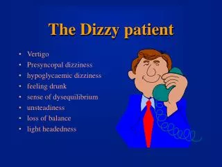
history • which complaints are related to vestibular deficits ? • patients often do not know which complaints are • associated with peripheral vestibular dysfunction • patients often think and are afraid that the complaints point • to a brain dysfunction • complaints are frequently a complex mixture of acute …. • and sustained symptoms !!!
history: my tip • when taking the history assume that there were and are • acute transient and sustained vestibular complaints • untill you find out that that is not the case
somatosensory foot sole pressure vision gravitoreceptors CNS interpretation learning adaptation compensation labyrinths hearing autonomic processes blood pressure heart beat frequency respiration rate circadian rhythm image stabilisation spatial orientation balance control
symptoms of vestibular dysfunction • acute loss or fluctuating peripheral vestibular function • transient: vertigo, nausea, falling / imbalance • remaining peripheral vestibular function loss • sustained: • - not feeling well, slight nausea • loss of balance at low speeds or complex situations • reduced dynamic visual acuity • reduced ability to discriminate between • self-motion and environmental motion • secondary: fear and fatigue
patient with severe bilateral vestibular hyporeflexia slow tandem walk fast tandem walk
cortex c mes cer cgl thal pons omn vn VOR: 8 msec OKR and Smooth pursuit: >75 msec
simulation of oscillopsia reduced dynamic visual acuity in case of bilateral vestibular areflexia
acute loss or fluctuating peripheral vestibular function • transient: vertigo, nausea, falling / imbalance • remaining peripheral vestibular function loss • sustained: • - not feeling well, slight nausea • loss of balance at low speeds or complex situations • reduced dynamic visual acuity • reduced ability to discriminate between • self-motion and environmental motion • secondary: fear and fatigue
5 major patterns Bronstein and Lempert ”Dizziness” • single episode of prolonged vertigo + sustained complaints • recurrent vertigo + sustained complaints • recurrent dizziness + sustained complaints • positional vertigo, less often sustained complaints • chronic dizziness, impaired visual acuity, unsteadiness
a vestibular function loss implies permanent impairment analogue to hearing and visual losses … and neuroplasticity differs per patient…!
bed-side examination • balance • observe patient at entrance • Romberg eo/ec, tandem walk slow vs fast • oculomotor • gaze and fixation • convergation / amblyopia / cover test / skew deviation • pursuit and saccades • static vestibulo-ocular stability • spontaneous nystagmus* • positioning • Hallpike AD/AS * + barbecue AD/AS * • VOR • head shake 3D VOR + OCR* • head shake nystagmus test* • head impulse test (H/V) • additional • fixation suppression test • test for fistula and Tullio phenomenon * preferrably with • Frenzels glasses
without Frenzel’s glasses • observe patient’s gait / posture • Romberg + tandem • if abnormal: past pointing test • 3. gaze and fixation • 4. convergence, amblyopia, • cover test, skew deviation • pursuit • saccades • with Frenzel’s glasses • 6. spontaneous nystagmus • 7. Hallpike + HC-test • 8. 3d VOR + OCR • 9. head shake nystagmus test • without Frenzel’s glasses • 10. head impulse test (H/V) • 11. fixation suppression test • 12. observe patient’s gait / posture specific bed-side examination of the vestibular function
spontaneous eyes open nystagmus vertical, horizontal symmetric or pendular always central (acquired or congenital) 1st, 2nd or 3rd degree horizontal mostly peripheral sometimes central
impact of visual fixation upon nystagmus nystagmus increases by visual fixation always central (acquired or congenital) nystagmus decreases upon visual fixation always peripheral
PC canalolithiasis or cupulolithiasis: most common peripheral vestibular dysfunction right left right PC-canalolithiasis or cupulolithiasis Hallpike left PC-canalolithiasis or cupulolithiasis Hallpike left or right AC canalolithiasis or cupulolithiasis Hallpike sidewards or mid-Hallpike right HC-canalolithiasis sidewards or mid-Hallpike left HC-canalolithiasis sidewards or mid-Hallpike right HC-cupulolithiasis left HC-cupulolithiasis sidewards or mid-Hallpike
right left right PC-canalolithiasis Hallpike left PC-canalolithiasis Hallpike left or right AC canalolithiasis or cupulolithiasis Hallpike right left sidewards or Hallpike right HC-canalolithiasis geotropic sidewards or Hallpike left HC-canalolithiasis geotropic sidewards or Hallpike right HC-cupulolithiasis apo-geotropic left HC-cupulolithiasis apo-geotropic sidewards or Hallpike
right left right PC-canalolithiasis Hallpike left PC-canalolithiasis Hallpike left or right AC canalolithiasis or cupulolithiasis Hallpike sidewards or mid-Hallpike right HC-canalolithiasis exclude neurological origin of a down beat nystagmus sidewards or mid-Hallpike left HC-canalolithiasis sidewards or mid-Hallpike right HC-cupulolithiasis left HC-cupulolithiasis sidewards or mid-Hallpike
normal tests: if history points to deficit manage patient in line with the history (but no ablative therapies)
optimal patient management: reality • a vestibular deficit implies permanent function loss • stimulation of neuroplasticity and use of rehabilitation exercises in natural environment improve function: • time is valuable: act fast • frequently only the history points to a vestibular deficit • explaining the relation between the deficit and the complaints forms the keystone of the therapy, • allowing the patient to cope with his or her problems