Download
1 / 75
1.71k likes | 3.68k Views
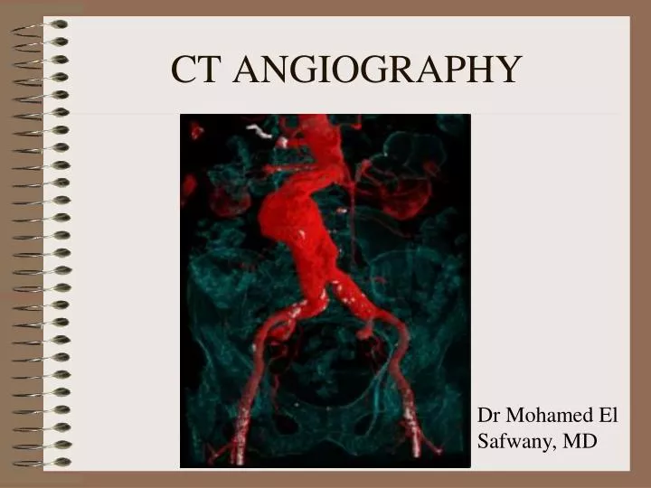
CT ANGIOGRAPHY. Dr Mohamed El Safwany, MD. Intended learning outcome. The student should learn at the end of this lecture CT IMAGE OF THE BLOOD VESSEL OPACIFIED BY CONTRAST. CT IMAGE OF THE BLOOD VESSELS OPACIFIED BY CONTRAST. REQUIREMENTS FOR CTA. PATIENT PREPARATION ACQUSITION PARAMETERS

E N D
CT ANGIOGRAPHY Dr Mohamed El Safwany, MD
Intended learning outcome The student should learn at the end of this lecture CT IMAGE OF THE BLOOD VESSEL OPACIFIED BY CONTRAST.
REQUIREMENTS FOR CTA • PATIENT PREPARATION • ACQUSITION PARAMETERS • CONTRAST MEDIUM ADMINISTRATION • POSTPROCESSING TECHNIQUES
PATIENT PREP. • CREAT.- 0.6 –1.2 mg/dl • IODINE ALLERGY- STEROID THERAPY • HYPERVENTILATION BEFORE EXAM FOR BETTER BREATHOLD
GAUGE SUITABLE FOR CTA 18 OR 20
PARAMETERS • USUALLY ROUTINE CT PRECEDES A CTA EXAM. THE ROUTINE EXAM IS USED AS A REFERENCE SCAN HELPING TO DETERMING THE SCANNING RANGE IN CTA.
SLICE THICKNESS SPATIAL RESOLUTION SLICE THICKNESS
CEREBRAL CTA ABDOMINAL CTA THORACIC CTA 1MM (LOWER mA) 3MM 3MM SLICE THICKNESS
PITCH SPATIAL RESOLUTION SPIRAL PITCH
SPIRAL PITCH • UP TO 2
kVp, mA, TIME • SIMILAR TO NON- CTA EXAM OF THE SAME BODY PART
RECONSTRUCTION INTERVAL • 50% OF OVERLAP
POWER INJECTOR PARAMETERS VOLUME OF CONTRAST-ml RATE ml/sec TIME OF INJECTION – sec SCAN DELAY TIME - sec
CONTRAST WARMER WHY???
RATE OF INJECTION 3-5 ml/sec
ORAL CONTRAST???? WATER NEGATIVE CONTRAST
AUTOMATED CONTRAST ADMINISTRATION SYSTEMS • SMARTPREP • CARE • SURESTART
CONTRAST ADMINISTRATION AND SCANNING METHODS • BOLUS TRACKING • BOLUS TIMING • MANUAL PRESET TIME
Circle of Willis?! 40 SEC
CERBRAL CTA CAROTID CTA CHEST CTA (PE) 15 SEC 12 SEC 10-20 SEC MANUAL DELAYS
TWO TECHNIQUES TO REDUCE MOTION ARTIFACTS IN CARDIAC CT • PROSPECTIVE TRIGGERING • RETROSPECTIVE GATING
Prospectively ECG-Triggered Sequential ScanningCardiac CT applications require the synchronization of data acquisition to the cardiac cycle, i.e. to the movement of the heart. For sequential imaging, a prospective trigger is derived from the ECG-trace to initiate the CT-scan with a certain delay time after the R-wave. Usually, the delay is defined such that the scans are acquired during the diastolic phase of the heart
Retrospectively ECG-Gated Spiral ScanningFor "retrospectively ECG-gated spiral scanning" a continuous spiral scan is acquired with the ECG-signal recorded simultaneously. The acquired scan data is selected for image reconstruction with respect to a pre-defined cardiac phase. Similar to ECG-triggered sequential scanning a certain R-wave delay time defines the start point of data that is used for image reconstruction. ECG-gated spiral scanning has several advantages over ECG-triggered sequential scanning. The continuous acquisition allows for reconstruction of overlapping slices. Due to the retrospective analysis of the ECG, the technique is less sensitive to arrhythmia.