Download
1 / 34
370 likes | 441 Views
This insightful chapter introduces the fundamentals of histology (the study of tissues) and embryology (the study of fetal development) in relation to human anatomy. Covering cell structure, tissue types, organ systems, and the process of embryonic development, it explains the significance of studying histology, essential techniques like microscopy and staining, and the relevance to medical education. Detailed information on common histological methods, tissue preparation for microscopy, and staining procedures are provided, with key points highlighted for easy comprehension. The chapter also emphasizes the importance of understanding the structure-function relationship in tissues and offers additional resources for further learning. Ideal for medical students, this resource-rich guide aims to enhance understanding and appreciation of histology and embryology concepts.

E N D
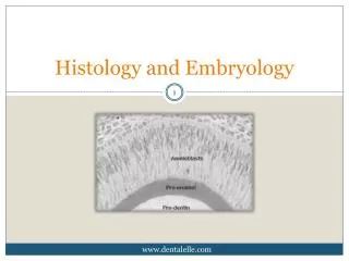
Histology and Embryology Zhong jie Li (李仲杰) School of medicine, Zhejiang University lizhongjie@zju.edu.cn
Relationship: cell --- tissue---organ---system---human body
Key points: I. What’s histology? II. Why we study it ? III. How to study it ? ---Histological methods.
I. What’s histology? Histology (Greek words): /histo---tissue /logia---study of ,or knowledge of Histology means the knowledge of tissue, is a branch of Anatomy.
Anatomy: ---gross anatomy ---microscopic anatomy/microanatomy Histology : a science which study the microstructure and the relationship between the structure and function of human being.
Cell: smallest unit of structure and function of body Tissue: group of cell and extracellular matrix Four basic tissue: ---Epithelial tissue ---Connective tissue ---Muscular tissue ---Nervous tissue
Organ: made up of tissue, have special shape, structure and function --- the cavity organ --- parenchymatous organ histology anatomy
System: organs which have related function get together.
What’s Embryology? Embryology is a kind of science which study the processes and the regulations of the development of human fetus. --- Preembryonic period --- Embryonic period (8 weeks before) --- Fetal period (9-38 weeks)
II. Why medical students should study Histology ?
Graduate Intern (exercitation) Clinic (Medicine,Surgery,Gynecology, Pedology, etc.) Pathphysiology Pharmocology Basic medicine &Surgery Physiology Biochemistry Microbiology Immunology Pathology Parasitology Histology & Embryology Anatomy
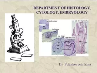
III. How to study it • Microscopy: Light microscopy (LM) Electron microscopy (EM) • Transmission electron microscope (TEM) • Scanning electron microscope (SEM) • Staining: H&E Histochemistry Immunohistochemistry In situ hybridiztion …
Relative Sizes in Histology Standard Units of Measure
Light Microscopy • :The resolution of the human eye is 0.2 mm. • :The resolution of the light microscope is 0.2 um. • :The resolution of the light microscope is 0.2 nm.
Preparation of tissue for LM The most routine one is paraffin section stained with hematoxylin and eosin(H&E) The steps: a.Obtaining the specimen: fresh, small pieces tissue block( less than 5mm3) b.Fixation: fixatives: use formalin or Bouin’s to preserve structural organization c.Dehydration: use ethyl alcohol to get rid of water of tissue and cell
d.Clearing: use xylene to get rid of alcohol *alcohol and xylene are embedding mediums e.Embedding: firstly, heat the paraffin, make it melt, then put tissue block into melted paraffin, allow paraffin harden, the tissue block is embedded in.
f.Sectioning: use microtome to cut the tissue into 3-8um thick sections, then mounted them on glass slides
H&E --- Routine staining for LM • ---Hematoxylin: basic dye, combines with acidic components, make them appear blue colour- basophilic, i.e. cell nucleus, hyaline cartilage • ---Eosin: acidic dye, combines with basic components, make them appear pink colour- acidophilic (eosinophilic), i.e. cytoplasm • basophilic • acidophilic (eosinopilic) • neutrophilc nucleolus nucleus
TEM: transmission electron microscopy The resolution of the TEM is about 0.1-0.5nm.
Basophilic granulocyte (TEM) Basophilic granulocyte (LM)
SEM: scanning electron microscopy The resolution of SEM is about 5nm.
Histochemistry & Cytochemistry To combine histological and cytological methods with chemical and biochemical methods and reveal the chemical composition of tissue and cell in situ.
Periodic acid-Schiff (PAS) reaction Localize polysaccharides, such as glycogen, in tissues and cells. PAS+ Produce an insoluble magenta complex
Immnohistochemistry • To use labelled antibodies as specific reagents for localizing tissue and cell constituents (antigens) in situ • Observation:Fluorescence microscope
In Situ Hybridization • A method for detection of specific RNA or DNA sequences directly in cells or tissue sections • incubate tissues with probe to detect cells expressing gene
Methods of learning: ---Must think in 3 dimensions ---Structure and function ---part and whole. oblique section Horizontal section longitudinal section
Structure and function Part and whole 舒张状态 收缩状态
Reference books • 1. Jun min T, Ji cheng L. Textbook of Histology and Embryology. Peking University Medical Press, 2011 • 2. William k. Ovalle & Patrick C. Nahirney. Netter’s essential histology, Elsevier Health Sciences,2007 • 3. Gaetner MP, Hlatt JL. Colour Textbook of Histology. Williams & Wilkins, 1997 Course web: http:/m-learning.zju.edu.cn (2015 Histology and Embryology)
The composition of final score: 1. Lecture and Lab attendence: 10 % 2. Quiz: 15% (each quiz 5%) 3. Lab test: 25% 4. Final written examination:50% Histology:35-40% Embryology:10-15% *. If you want to pass this course, final written examination score must be more than 50. Preparation of laboratory work • Have the tools ready: • Pencils (red-blue pencil) • rubber, ruler and so on