Download
1 / 14
190 likes | 417 Views
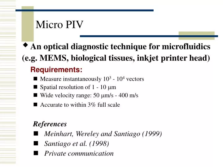
Micro PIV. An optical diagnostic technique for microfluidics (e.g. MEMS, biological tissues, inkjet printer head) Requirements: Measure instantaneously 10 3 - 10 4 vectors Spatial resolution of 1 - 10 m m Wide velocity range: 50 m m/s - 400 m/s Accurate to within 3% full scale

E N D
Micro PIV • An optical diagnostic technique for microfluidics (e.g. MEMS, biological tissues, inkjet printer head) Requirements: • Measure instantaneously 103 - 104 vectors • Spatial resolution of 1 - 10 mm • Wide velocity range: 50 mm/s - 400 m/s • Accurate to within 3% full scale References • Meinhart, Wereley and Santiago (1999) • Santiago et al. (1998) • Private communication
Video Microscopy • Mature technology in bio-medical fields The smallest resolvable size dp = l/NA , NA (Numerical Aperture)= n sinq For comparison, recall diffraction limit for camera: ddiff = 2.44l/(D/f)=2.44l(f#) • Microscopy + PIV Resolve particles of sub-microns Measurement of particle displacement Image field: 30~300mm n q dp
Field of View: 30 ~ 300 mm Vector Spacing: 1 ~ 10 mm Interrogation Cell: 2 ~ 20 mm (50 % overlap) min. 10 pairs of particles for correlation “Plane” Thickness dz: Depth of Field of microscope ~ 1mm 30 ~ 300 mm 1 ~ 10 mm 2 ~ 20 mm Laser sheet thickness ~ 1 mm Micro PIV vs. PIV Shrink 1000 times
Micro PIV Small-- Follow flow Do not clog the device Do not alter fluid property But not too small-- Suppress Brownian motion Generate enough light signal Dp = 0.3 ~ 0.7 mm Regular PIV Small enough to track flow, need to be detectable by the camera Dp = 3 ~ 30 mm Tracer Particles
Challenges by Sub-micron Particles • 1. Optical Resolution: need Dp = 300 – 700 nm (Nd:YAG: l ~ 500 nm) Visible light400 nm-----750 nm If NA <1, cannot resolve dp less than l sin q <1 n: index of refraction between specimen & objective • 2. Low Light Signal
Solutions • Oil immersion lens (n 1.5) to get NA >1 NA =1.4for 60x - 100x objectives • Fluorescence (epi-illumination, reflection) dp < l & stronger signal • Differential Interference Contrast (DIC) microscopy Shearing interference to highlight refraction change
Mercury arc lamp Exposure t ~ 2 ms Pulse delay Dt ~ 100 ms (Also depend on camera transfer) Velocity up to 50 mm/s Pulsed laser (Dual Nd:YAG laser) t ~ 5 ns Dt ~ 500 ns up to 1 m/s Light Source and Camera Digital CCD Camera (1030 x 1300 x 12 bit cooled interlined transfer can record back-to-back images within 500 ns)
Data Processing • Correlation • Significant Noise: • Out-of-plane motion • Brownian motion • Ensemble-averaging correlation technique (average 20 instantaneous correlations) • Limited to steady or periodic flows
Example 1 • Santiago et al. (1998)
Result • Santiago et al. (1998)
Meinhart, Wereley and Santiago (1999) Example 2
Meinhart, Wereley and Santiago (1999) Result Ensemble-averaged velocity-vector field measured in a 30 mm deep, 300 mm wide, 25 mm channel. The spatial resolution is 13.6 mm x 4.4 mm away from the wall, and 13.6 mm x 0.9 mm near the wall. A 50% overlap between interrogation spots yields a velocity vector spacing of 450 nm in the wall- normal direction near the wall
Inkjet Printer Head • Field of view 50 ~ 500 mm • Need objective lens working distance >1mm (Cover Glass) Smaller NA Larger particle size (~ 0.6) (~ 0.7 mm) • Unsteady flow in the cycle of droplet ejection: need instantaneous or phase-averaged measurement
Basic Limitation of Micro PIV • DOF (~ 1mm) limits to strictly 2D flow • Not only 2D vector map, • Out-of-plane motion can cause measurement to fail • Hence must select a plane with only 2D motion PIV Plane