Download
1 / 46
460 likes | 725 Views
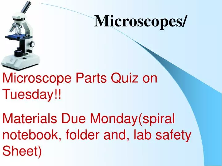
Microscopes/. Microscope Parts Quiz on Tuesday!! Materials Due Monday(spiral notebook, folder and, lab safety Sheet). It is estimated that the human race grows daily by about 214,000 people.

E N D
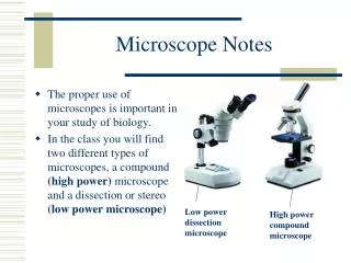
Microscopes/ Microscope Parts Quiz on Tuesday!! Materials Due Monday(spiral notebook, folder and, lab safety Sheet)
It is estimated that the human race grows daily by about 214,000 people. • It takes only 15 watts of electricity going through a human body to stop the heart. Common lightbulbs run on about 25 to 75 watts of electricity.
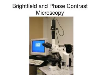
Microscopes • Objectives: • To name the parts of the microscope and describe their functions
Microscopes • Objectives: • To name the parts of the microscope and describe their functions • To describe how to properly use a compound microscope.
Microscopes • Objectives: • To name the parts of the microscope and describe their functions • To describe how to properly use a compound microscope. • To compare a compound to a light microscope
Label the parts on your microscope picture. Eyepiece/ocularlensmagnification = 10X
Label the parts on your microscope picture. Eyepiece/ocularlensmagnification = 10X Arm-Support
Label the parts on your microscope picture. Eyepiece/ocularlensmagnification = 10X Arm-Support Stage - support
Label the parts on your microscope picture. Eyepiece/ocularlensmagnification = 10X Arm-Support Stage - support Coarse adjust – general focus
Label the parts on your microscope picture. Eyepiece/ocularlensmagnification = 10X Arm-Support Stage - support Coarse adjust – general focus Fine Focus –high power focus
Label the parts on your microscope picture. Eyepiece/ocularlensmagnification = 10X Arm-Support Stage - support Coarse adjust – general focus Fine Focus –high power focus Base -support
Label the parts on your microscope picture. Eyepiece/ocularlensmagnification = 10X Arm-Support Stage - support Coarse adjust – general focus Fine Focus –high power focus Light source Base -support
Label the parts on your microscope picture. Eyepiece/ocularlensmagnification = 10X Arm-Support Stage - support Coarse adjust – general focus Diaphragm – adjusts amount of light Fine Focus –high power focus Light source Base -support
Label the parts on your microscope picture. Eyepiece/ocularlensmagnification = 10X Arm-Support Stage - support Stage clips – holds slide Coarse adjust – general focus Diaphragm – adjusts amount of light Fine Focus –high power focus Light source Base -support
Label the parts on your microscope picture. Eyepiece/ocularlensmagnification = 10X Arm-Support Objective Lenses Scanning = 4X Low Power = 10X High power = 40X Stage - support Stage clips – holds slide Coarse adjust – general focus Diaphragm – adjusts amount of light Fine Focus –high power focus Light source Base -support
Label the parts on your microscope picture. Eyepiece/ocularlensmagnification = 10X Revolving nosepiece Arm-Support Objective Lenses Scanning = 4X Low Power = 10X High power = 40X Stage - support Stage clips – holds slide Coarse adjust – general focus Diaphragm – adjusts amount of light Fine Focus –high power focus Light source Base -support
Label the parts on your microscope picture. Eyepiece/ocularlensmagnification = 10X Revolving nosepiece Arm-Support Objective Lenses Scanning = 4X Low Power = 10X High power = 40X Stage - support Stage clips – holds slide Coarse adjust – general focus Diaphragm – adjusts amount of light Fine Focus –high power focus Light source Base -support
Eyepiece Objective Lens Magnification Scanning lens (red) 10X Low Power (yellow) 10X High Power (blue) 10X • Magnification of a Compound Microscope • Because you are looking through multiple lenses the lenses have a “compounding” effect. • The eyepiece always magnifies 10X
Eyepiece Objective Lens Magnification Scanning lens (red) 10X 4X Low Power (yellow) 10X 10X High Power (blue) 10X 40X • Magnification of a Compound Microscope • Each of the objective lenses have their own magnification
Eyepiece Objective Lens Magnification Scanning lens (red) 10X 4X Low Power (yellow) 10X 10X 100X High Power (blue) 10X 40X 400X • Magnification of a Compound Microscope • You then multiply the eyepiece with the objective lens to determine the total magnification 40X
Other Laboratory Techniques • Staining • Dyes are added to slides to bring out detail and stain certain features. • Centrifugation • A device that can spin tubes up to 20,000X/min. This is used to separate samples (I.e. parts of cells • Cell Culture • Cells of a particular kind are grown on plates with all their required nutrients.
As we focus the microscope to higher powers we see more and more detail, until a certain point. Then objects become blurry and detail is lost. Think of blowing up a picture too big. • Resolution is out ability to distinguish two points as separate. • Electron Microscopes have a much higher limit of resolution. Limits of Resolution Click here to zoom
The Electron MicroscopeAllows us to see very high resolution images SCALES AND SETAE ON LEPIDOPTERA Dust mite PREDATORY ANT WITH PEAR PSYLLA IN ITS MOUTH
The Electron MicroscopeHow does it work? They use giant electromagnets to sent a stream of electrons over the specimen. This image is then read by a computer.
General Procedures • ·Make sure all backpacks are out of the aisles before you get a microscope! Always carry the microscope with one hand on the Arm and one hand on the Base. • ·Wear your glasses, the microscope will focus to your eyesight! • ·Keep both eyes open, your brain will learn to ignore the other eye.
Focusing a Specimen • Always start on low power or scanning.
Focusing a Specimen • Always start on low power or scanning. • Focus the specimen using the course adjust.
Focusing a Specimen • Always start on low power or scanning. • Focus the specimen using the course adjust. • Re-center. If high power is needed, turn only after re-centering!
Focusing a Specimen • Always start on low power or scanning. • Focus the specimen using the course adjust. • Re-center. If high power is needed, turn only after re-centering! • Now use the FINE FOCUS ONLY for your final adjustments.
Making a Wet Mount 1. Gather a very thinslice/piece of whatever your specimen is. All samples should be paper thickness or thinner! 2. Place ONE drop of water directly over the specimen. 3. Place the cover slip at a 45 degree angle with one edge touching the water drop, and let go.
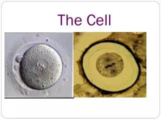
Making good drawings • Don’t even think of starting your drawing unless you have a pencil (colored are even better) drawings in pen are unacceptable.This is for two reasons: (a) You can erase pencil! (b) You can shade in areas more easily in pencil • In the upper left hand corner of each circle include the specimen name as written on the slide label. In the upper right hand corner, include the magnification (100x or 430x). • Label Everything you identify! Cheek cell 100X Cell membrane nucleus
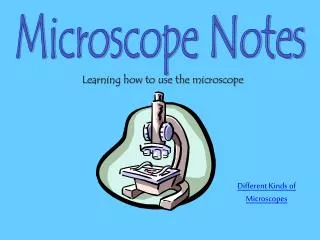
Student handouts • Microscope notes page
Microscopes Eyepiece Objective Lens Magnification Scanning lens (red) 10X Low Power (yellow) 10X High Power (blue) 10X Microscope Parts Label with structure and function Magnification of Lenses
Light verses Electron Microscope Practice live Cell Sketches