Download

1 / 60
600 likes | 817 Views
Kidney & Urinary Tract Neoplasms. Jaroslava Dušková. Kidney Cancer. 2% of the total human cancer burden, M:F 2:1, middle age preference for developed (industrialized) countries risk factors: TOBACCO SMOKING, OBESITY. silent for a long time

E N D
Kidney & Urinary Tract Neoplasms Jaroslava Dušková
Kidney Cancer • 2% of the total human cancer burden, M:F 2:1, middle age • preference for developed (industrialized) countries • risk factors: TOBACCO SMOKING, OBESITY
silent for a long time - discovered by chance hematuria, backache, abdominal mass, metastatic spread early hematogenic spread possible Symptoms
WHO classification of tumours of the kidney (2004)
Renal cell (12) Metanephric (3) Nephroblastic (3) Mesenchymal (18) Mixed mesenchymal and epithelial (3) Neuroendocrine (5) Hematopopietic and lymphoid (3) Germ cell (2) Metastatic (-) WHO Histogenetic groups (& number of nosology units identified)
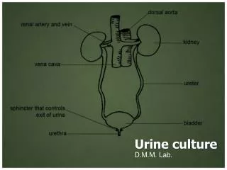
Epithelial Neoplasms of the Pelvis • Benign - papillomas • Malignant - carcinomas • papillocarcinomas • squamous cell Urinary ways
Benign Malignant Kidney Tumours
Kidney Adenoma Definition: • Formerly - diam. 2-3 cm • Recently – only diam. less than 5mm without a clear cell component • tubulopapillary architecture • lack of atypiae & mitoses
benign ADENOMAS papillary tubulopapillary (<5mm!) oncocytic (oncocytoma) metanephric Epithelial Kidney Tumours
Oncocytoma • Kidney cortex • may be multicentric and bilateral • Macro – tan with a central stellate scar • Micro - eosinophillic granular cytoplasm bizarre nuclei • Elmi – mitochondria filling up the cytoplasm • Biological behaviour benign
Angiolipoleiomyoma – mixed mesenchymal tumour Kidney Tumours - mesenchymal
Metanephric Adenoma • small dark cells • acinar and glomeruloid formations • calkospherites, calcifying non agressive
Benign Kidney Tumours Mimicking Carcinomas and Sarcomas • Metanephric adenoma - large & cellular • Oncocytoma - large with atypiae • Angioleiomyolipoma - large with atypiae
malignant CARCINOMAS Clear Conventional Cell Papillary (chromophillic) type 1 type 2 Chromophobe classical eosinophillic Sarcomatoid Cystic Collecting Duct Epithelial Kidney Tumours
Clear Cell Ca (Grawitz tumour) (75%) • Solid / cystic • Unilocullar or multilocular • Micro - solid or tubulocystic clear cytoplasm (fat & glycogen) • Immunohistochemistry cytokeratins, vimentin, CD10, EMA, S-100 • Cytogeneticsdeletion of the short arm chromosome 3 (3p) Prognosis: G, pT dependent Sarcomatoid variant is the most malignant
Papillary (Chromophillic) Ca (10%) • In dialysed more frequent • X-ray hypovascular • Histology –papillary/ tubulopapillary type 1 – cubic cells type 2 - cylindric cells (worse prognosis) • Genetics –trisomy ortetrasomy 7 and 17 in men often Y chromosome missing mutation of c-met oncogen Prognosis : G, pT dependent slightly better than in conventional ca
Chromophobe Carcinoma (5%) • Macro - brown color • Mikro - solid, cytoplasms clear or eosinophillic, positive in Hale´s colloidal iron staining, raisin-like cell nuclei • Elmi microvesicles in cytoplasm • Genetics missing chromosomes - 1, 2, 10, 13, 6, 21, 17 Prognosis: G, pT dependent
Collecting Duct Carcinoma • Starts in the medulla • Micro • adenocarcinoma & urothelial like • hobnail cells • papillary • fibroplasia, mucin production • Imunocytokeratin 13, vimentin, lectin Prognosis unfavourable
Nephroblastoma (Wilms´tumour) • syn. - embryonal adenosarcoma • Children - preschool age • Macro: gray-white large retroperitoneal mass palpable through abdominal wall • Micro: undifferentiated renal blastema, tubular and glomeruloid formations may be present • Prognosis: curable (stage!) • Follow up: - nephroblastomatosis
Role of the Pathologist in the Kidney Tumour Diagnostics • Typing • Biological Behaviour • Grading • Staging
Grading • Nuclear – Fuhrman et al. 1982 • Nuclear plus architecture • Proliferation factors - PCNA, Ki 67, Bcl 2 • Morphometry • DNA Analysis • AgNOR • Angiogenesis • Cytometry Flow cytometry
Staging • Size • Kidney capsule infiltration • Angioinvasion • Metastases in the lymph nodes • Number of lymph nodes involved • Metastases in the surrounding organs
Nuclear Grading in Kidney Cancer (Fuhrman et al. 1982) • Grade I small, uniform, round (10 ) inaparent or missing nucleoli • Grade II larger irregular (15 ) nucleoli small • Grade III large, irregular margins (20 )nucleoli large • Grade IV large, bizarre, pleomorphic
Factors with an Adverse Prognosis Influence in Kidney Cancer Sizediam. more than 12 cm Invasion to venes recidives GradingG III and G IV Staging most important Proliferation Index p53Expression
Kidney Cancer – complications 1. • metastatic spread & generalisation • manifestation via solitary bloodborne metastasis possible (pathological fracture, struma neoplastica…) • hematuria – anemia
Kidney Cancer – complications 2. • hormon production – erythropoietin polyglobulia Wood L, Swanepoel C, du Toit A, Jacobs P.Clinically silent renal tumour producing erythropoietin.S Afr Med J. 2003 Feb;93(2):128-9. Shaheen M, Hilgarth KA, Hawes D, Badve S, Antony AC.A Mexican man with "too much blood". Lancet. 2003 Sep 6;362(9386):806. • insulin, glukagon, renin, HPL like substances
Urothelial Cancer • approx. 3% of total human cancer burden • increasing incidence • industrialized countries • risk factors: TOBACCO SMOKING aniline dye industry phenacetin schistosomiasis
Symptoms hematuria (obstruction) (metastases)
Terminology …the term UROTHELIAL be used rather than „transitional“...
Normal urothelium multilayered variable number of layers empty bladder 4 - 6 full bladder 2 - 3
Normal urothelium Cells: basal superficial („umbrella“) polyploid, binuclear neuroendocrine
„Variations“ of Urothelium – slight reactive changes von Brunn´s nests mucinous metaplasia squamous metaplasia (nonkeratinising, vagina type)
Metaplasia Def:change of one differentiated structure into another one (e.g. urothelium – squamous epithelium)
Cause: iritation Types: squamous nonkeratinizing keratinizing mucinous nephrogenic clear cell Urothelium Metaplasia
Metaplasia Significance: • dif. dg. problem • with atypia precancerosis
Submucose discontinual muscularis mucosae continual row of vessels important for staging of urothelial ca (pT1a, pT1b, pTx)
The WHO/ISUP Consensus Classification of Urothelial Neoplasmsof the Urinary Bladder Epstein JI, Amin MB,Reuter VR, Mostofi FK, & the Bladder Consensus Conference Committee Am.J. Surg. Pathol.,22,1998,1435-8 WHO 2004
The WHO/ISUP Consensus Classification • Hyperplasia • Flat lesions with atypia • Papillary neoplasms • Invasive neoplasms
The WHO/ISUP Consensus Classification I. Hyperplasia Flat Papillary
Hyperplasia Def:regular increase in number of uroth. layers (min. >7, mostly >10) slight increase in cell nuclei size, preserved architecture
Hyperplasia Significance: precancerosis 70% of patients with urothelial ca identical mutations
The WHO/ISUP Consensus Classification • Hyperplasia • Flat lesions with atypia • Papillary neoplasms • Invasive neoplasms
II. Flat lesions with atypia • Reactive (inflammatory) atypia • Atypia of unknown significance • Dysplasia (LG IUN) • CIS (HG IUN)
Atypia of uncertain significance Def.: urothelial changes similar to reactive (inflammatory) ones where anusually high intensity of atypiae compared to minimal inflammatory background is present
Dysplasia DEF: disturbance of normal urothelium architecture & cytology
Dysplasia • with an inflammatory background • without -“- in a flat urothelium in the papillary urothelium
Dysplasia LG IUN – low grade intraurothelial neoplasia HG IUN/ CIS – high grade intraurothelial neoplasia
The WHO/ISUP Consensus Classification • Hyperplasia • Flat lesions with atypia • Papillary neoplasms • Invasive neoplasms
III. Papillary neoplasms • Papilloma • Inverted papilloma • Papillary Urothelial Neoplasm of LowMalignant Potential PUNLMP • Papillary carcinoma, low grade • Papillary carcinoma, high grade