Download
1 / 27
270 likes | 511 Views
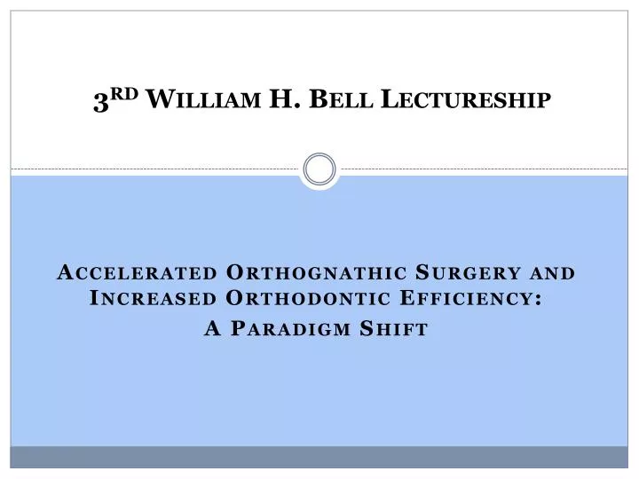
3 rd William H. Bell Lectureship. Accelerated Orthognathic Surgery and Increased Orthodontic Efficiency: A Paradigm Shift. Volumetric Three-Dimensional Upper Airway Analysis in Patients with Obstructive Sleep Apnea Following Maxillomandibular Advancement.

E N D
3rd William H. Bell Lectureship Accelerated Orthognathic Surgery and Increased Orthodontic Efficiency: A Paradigm Shift
Volumetric Three-Dimensional Upper Airway Analysis in Patients with Obstructive Sleep Apnea Following Maxillomandibular Advancement Joseph A. Broujerdi, MD, DMD Richard L. Jacobson, DMD, MS Stephen A. Schendel, MD, DDS, FACS Prepared by Jenny R. Armstrong
References • Maxillomandibular advancement is an effective treatment option for patients with sleep apnea • Holty JE, Guilleminnault C. Maxillomandibular Advancement for the Treatment of OSA: A Systematic Review and Meta-Analysis, Sleep Medical Review. 2010 Oct.; 14(5):287-97 • Schendel S., Powell N., Jacobson R. Maxillary, Mandibular, and Chin Advancement: Treatment Planning Based on Airway Anatomy in Obstructive Sleep Apnea. Journal of Oral and Maxillofacial Surgery. 2011 March; 69(3):663-76 • 3-Dimensional volumetric analysis is reproducible and accurate • Schendel S. Automated 3-D Airway Analysis by Cone Beam CT. Journal of Oral and Maxillofacial Surgery. 2010 March; 68(3):696-701 • KabanLB et al. Three-Dimensional Computed Tomographic Airway Analysis of Patients with Obstructive Sleep Apnea Treated by Maxillomandibular Advancement. Journal of Oral and Maxillofacial Surgery. 2011 March; 69(3):677-86
Surgical Technique • LeFort I Maxillary Osteotomy Advancement • Bone grafting • +/- Septoplasty and/or turbinectomy • Bilateral Sagittal Split Osteotomy of the Mandible Advancement • +/- Genioglossal Advancement
Pre- and Post- Op Data: Movement (mm) • Average Mandibular Movement: 9.57 mm • Average Maxillary Movement: 9.86 mm • Average Genio Movement: 6 mm
Pre- & Post- Operative Volumetric Analysis • Average % Change UAS • 236.99% • Average % Change RP • 361.97% • Average % Change RG • 164.56%
Pre- & Post- Operative Surface Area Analysis • Average Choke Point RP Pre • 78.04 mm2 • Average Choke Point RP Post • 237.45 mm2 • Average % Change RP • 664.22% • Average Choke Point RG Pre • 84.80 mm2 • Average Choke Point RG Post • 154.55 mm2 • Average % Change RG • 100.98% • Location of Choke Pt Pre-Operatively • 5 Patients Retropalatal, 2 Patients Retroglossal • Location of Choke Pt Post-Operatively • 6 Patients Retroglossal, 1 Patient retropalatal
Pre- & Post- Operative Transverse Analysis • Average change in the transverse dimension • Retropalatal: 11.64 mm • Retroglossal: 7.11 mm
Pre- & Post- Operative A-P Analysis • Average change in the A-P dimension • Retropalatal: 6.40 mm • Retroglossal: 2.61 mm
Pre- & Post- Operative Height Analysis • Average Change in UAS height • 2.86 mm • Average Change in RP height • 3.14 mm • Average Change in RG height • 4.29 mm
Pouseille’s Law As radius increases and height decreases, the resistance of flow decreases
Results • Volume • The UAS enlarged significantly • The shape of the UAS changed from a funnel to a tube like shape • The retropalatal space increases in volume more than the retroglossal space • Surface Area • The surface area at the choke point in the retropalatal space increases by a greater percent change than the retroglossal space • The location of the choke point is generally pre-operatively in the retropalatal space and post-operatively in the retroglossal space • Indication of normalizing the airway and eliminating any bottlenecking/funneling • The airway enlarges in a rectangular fashion • Length • The transverse dimension increases more than the A-P dimension in millimeter change • The A-P dimension increases more than the transverse dimension in percent change • The retropalatal space increases more in the transverse and A-P dimensions than the retroglossal space does • Height • The height of the airway measured from the posterior of the post nasal spine to the superior tip of the hyoid bone generally decreases post-operatively • The height of the airway was pre-operatively shorter in the retropalatal space than the height of the retroglossal space • The height of the airway was post-operatively shorter in the retroglossal space than the retropalatal space
Case I Pre-Surgical Post-Surgical
Case I Pre-Surgical Post-Surgical
Conclusions • 3-Dimensional airway analysis indicates that maxillomandibular advancement is an effective treatment option for patients with obstructive sleep apnea by increasing tension and changing the position of the palatal and pharyngeal muscles. As a result: • Airway volume increases • Shape of airway changes • Change from a funnel to cylindrical shape • Height of airway decreases • Resistance decreases • Radius of the airway increases • Height of the airway decreases
References • Maxillomandibular advancement is an effective treatment option for patients with sleep apnea • Holty JE, Guilleminnault C. Maxillomandibular Advancement for the Treatment of OSA: A Systematic Review and Meta-Analysis, Sleep Medical Review. 2010 Oct.; 14(5):287-97 • Schendel S., Powell N., Jacobson R. Maxillary, Mandibular, and Chin Advancement: Treatment Planning Based on Airway Anatomy in Obstructive Sleep Apnea. Journal of Oral and Maxillofacial Surgery. 2011 March; 69(3):663-76 • 3-Dimensional volumetric analysis is reproducible and accurate • Schendel S. Automated 3-D Airway Analysis by Cone Beam CT. Journal of Oral and Maxillofacial Surgery. 2010 March; 68(3):696-701 • KabanLB et al. Three-Dimensional Computed Tomographic Airway Analysis of Patients with Obstructive Sleep Apnea Treated by Maxillomandibular Advancement. Journal of Oral and Maxillofacial Surgery. 2011 March; 69(3):677-86