Download
1 / 33
330 likes | 342 Views
). EXCRITARY SYSTEM. DEPARTMENT OF PHYSIOLOGY.

E N D
) EXCRITARY SYSTEM DEPARTMENT OF PHYSIOLOGY
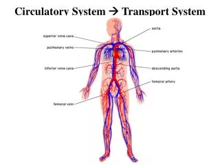
INTRODUCTION:-excretion is the process by which the unwanted substance & matabolic wastes are eliminated from the body. There are many organ & system involved in excritary function such as:- Kidney, skin, lungs, Gastro Intestinal Tract, Salivary Gland & Liver are main channels through which excretion takes place. There seems to be a general rule regarding the channel of excretion of different substances. For instance,
(1) The soluble non-irritant, solid substance & water mainly excreted through the kidneys & to a lesser extent through skin. (2) voletile substance such as Carbon-di-oxide, ammonia, alcohal, ketone bodies, aromatic oils, water vapours etc are excreted through lungs. (3) Heavy metals are excreted through G.I.T. especially through the large intenstine & slightly though the liver & saliva.
SYSTEM URINARY
(4) fats & fat darivatives are pased out of the body through the skin, as sebum & through the liver along with bile. These are the broad principles on which excretion from the body takes place. To understand the way in which the kidney carries out this function, it is esential to understand first the way in which it is supplied with blood.
KIDNEY:- INTRODUTION OF KIDNEY:-The human kidneys are bean-shaped paired organs situated juste behind the vertebral column in the abdomen at the T12 to L3 segment. An avrage sized kidney measures 10-12 cm in lengh, 5-6 cm in width & 3-4 cm in thickness, each weighing about 150 grm in adult male & about 135 grm in adult female. Usually the ritht kidney is slightly smaller than the left one. A deep notch “or” concavity is present at the medial border the hilus (hilum) & it is through this region the blood vessels- renal artary & vein, ureter & nerves pass.
STRUCTURE:- GROSS STRUCTURE:- (1) mainly section of kidney- (i) outer cortex- reddish in colour (ii) inner medulla- paler in colour (2) The ureters exit from the hilus of the kidney & pass to the bladder. The blood vessels, lymphatics and nerver enter into or exit from the kidney via the hilus.
MICROSCOPIC STRUCTURE:- The basic fuctional unit of kidney is nephron. There are aproximately 1-1.3 million nephrons in each kidney which drain into the renal pelvis.Total lengh of nephron ranges from 45-65 mm. (a) Bowman’s Capsule:- the initial dilate part of the nephron. It’s epithilial cells lining is about 5μm thick. (b)Glomerulus:-it is about 200 µm in diameter & form by the, invagination of a tuft of capillaries into bowman’s capsule. The capillaries are supplied by afferent arteriole & the blood leaves from the tuft efferent arteriole. Bowman’s capsule & Glomarulus togher costitute the Malpigian Carpsule.
The Bowman’s capsule has 2 layers (1) visceral cell layer & (2) parietal cell layer A space present between the visceral & parietal layers of bowman’s capsule called “Bowman’s Space”. The structures intervening the blood with the capillary loop and bowman’s space called “Glomerular Membrane” Or“Glomerula Capillary wall”.
STRUCTURE OF GLOMERULAR MEMBRANE:- 1. It is an extremely thin membrane and is made of 5 layers. (i) Foot processes of podocytes. (ii) Lamina rara externa or outer cement layer. (iii) Lamina densa. (iv) Lamina rara interna Or inner cement layer. (v) Endothelial cell layer. 2. The glomerular capillaries from a freely branching anatmosis network. Each glomerulus contains 6 lobules and each of these consists of 3-6 capillaries lobes.
Many anastomoses occur between the capillaries with in any one lobule. 3.The major function of glomerular membrane is to produce an ultrafiltrate. The glomerular filtrate will contain all the constituents of plasma except proteins. funtionally, The glomerular membrane permits the free passage of neutral substances upto 4 nm in diameter and almost totally exclusdes those with diamiter greater than 8 nm.
(c) PROXIMAL CONVULATED(PCT):- features:- 1.Pct luwen is continuous with that of Bowmen’s capsule. 2.It is 15 mm long and 55µm in diamiter consisting of single layer of cells with curved outline & brush border formed by numerous ‘microvilli’ which markedly increase the surface area for absorption.
3. Tubule cell are rich in mitocondria since they are responsible for the active active transport of about 80% of the sodium filtrated of the tubuler fluid into the perticuler capillary blood, There for corespondingly have a high role of oxygen consumption. 4. Tuble cells are united at the apex by tight juctions while the bases of the cells have extensions into extracellular spaces. 5. PCT is divided into two parts (i) pars convuluta - convoluted protion of PCT (ii) pars erecta - straight portion of PCT
(D) LOOP OF HENLE:- (1) It consist of a descending limb which arises in continuity with the terminal part of the PCT. (2) Descending limb continues into the thin segment where epithilium is of attenuated flat cell. Length of the thin segment of the loop is 2 to 14 mm. (3) From this segment arises the thick ascending limb ( lengh 12 mm) which is formed by low cuboidal epithelium. The cell of the ascending limb have numerous mitocondria and the basilar portion of their cell membrane are exhensively invaginated.
(E) DISTAL CONVULATED TUBULE (D.C.T.):-Thick ascending as DCT (length 5 mm). (1) DCT is characterized by low cuboidal epithilium of with few scattered microvilli. (2) This tubule comes very close to its own glomerulus and establishes a close proximily to the afferent and effrent arterioles of the glomerulus. At this site the cells of DCT get modified to become columnar and are closely crowded together, that is why this part of DCT is call “MACULA DENCA”.
(3) The macula densa and the adjucent Juxtra glomerula part of the affrent arterilar wall are functionally associated forming Juxta Glomerlar Apparatus (JGA).
(F) COLLECTION TUBULE (C.T.):- (1) DCT join to form Collecting tubules (Length 20 mm) and is lined by clear cuboidal epithelium. (2) CT passes through the renal cortex and medula to empty into the plevis of the kidney at the apices of the medullary pyramids. (3) CT epithelium consists of two types of cells :- (i) p-cells (Principal cells) which increase the permeability of CT to water in the presence of ADH. (ii) I-cells (Intercaluted cell) which secrete acid and are responsible for acidic urin.
The whole kidney is enveloped by a thin but tough fibrous membrane, called Renal Capsule. It limits the swelling if the kidney becomes oedelous this increases glomerular filtration rate (GFR) and precipitates Anuria. TYPES OF NEPHRON :- (1) Cortical nephrons or superficial nephrons (2) Juxtramedullary nephrons (1) Cortical nephrons :- The nephrons, which have their corpuscles in the outer cortex of the kidney near the periphery are called cortical nephrons are cortical nephrons.
(2) Juxtramedullary nephrons :- The nephrons which have their cortex near medulla or corticomedullary junction are called Juxtramedullary nephrons.
JUXTRA GLOMERULAR APPRATUS(JGA) The JGA is a combination of specialized tubular and vascular cello located at the vascular pale where the afferent and efferent arterioles enter and leave the glomerulus. It is composed of three cell type. 1. Juxtra glomerular cell (JG cell) 2. Macula Densa cell 3. Mesangial cells or Lacis cells
Regulation of Renin Secrition :- ٭Inhibition :- (1) Inhibition of cells by – (I) Stretch due to increase afferent arteriolar pressure (II) Angiotensin II (III) Vasopressin (ADH) (2) Increased NA+ or Clֿ reabsorption across macula densa.
*Stimulation :- (1) Stimulation of JG cells by – (I) increase in smpathetic activity via rena nerves(hypovalemia, hypotention) (II) increse circulating catecholamines (III) prostaglandins (2) Sodium depletion Diuretic Congestive Cardiac failure.
RENIN - ANGIOTENSIS SYSTEM :- The renin and angiotensin system is the key regulator of aldosterone release .the m- RNA for renin has been found in a number of tissues including brain, adrenal gland , uterus & placenta. It is believed that kidney is the physiologically most important site of renin synthesis. In kidney, renin is synthesized & storeg as a preproharmone in the cells of the Juxtraglomerular complex.
Renin release is stimulated by a number of factors which are influenced by changes in sodium load and plasma volume and include reduced sodium intake, decreased arterial pressure, a fall in extracellular fluid volume stress and trauma. Renin is synthesized and secreted by modified Smooth muscle cell in the walls of arterials when deacrese in ECF volumn is sensed by the kidney or decreased perfusion pressure detected by barorecepter in the afferent arterials resulting in increased Renin synthesis and release. Renin cause circulating angiotensinogen to be cleaved resulting in angiotensin – angiotensin I angiotensin II, the most patent vasoconstrictor. The enzyme catalizing this seaction is known as converting enzyme located primarly in the lung vascolature
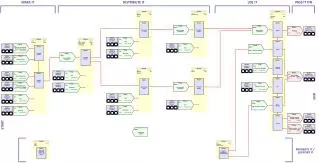
PRODUCTION OF ANDIOTENSIN II :- Angiotensinogen in plasma Fall in fall in renal increased B/d volu. perfusion renin(IGA) Angio. I Increased Aldosterone adrenal b/d volu. cortex Angio. II Inceased ADH kidney tubule sweat in lungs glands & salivary glands increase plasma osmotic pressure increase Na+,Cl-Arteriolar & H2o reabsoption vasoconstriction increased B.P. PRODUCTION OF ANDIOTENSIN II :- Angiotensinogen in plasma Fall in fall in renal increased B/d volu. perfusion renin(IGA) Angio. I Increased Aldosterone adrenal b/d volu. cortex Angio. II Inceased ADH kidney tubule sweat in lungs glands & salivary glands increase plasma osmotic pressure increase Na+,Cl-Arteriolar & H2o reabsoption vasoconstriction increased B.P.
HARMONES THAT CONTROL NaCl & WATER REABSOPTION :- “Proximal * Angiotensin II NaCl H2o Tubule” * Gluvovorticoids NaCl H2o “Thick * Aldosterone NaCl Ascending * Vasopressin NaCl Limb” “Distal * Aldosterone NaCl Convolated * Atrial Natiuretic peptide Nacl H2o And * Prostaglandins Nacl H2o Collecting * Bradykinin Nacl H2o Duct” * Vasopressin Nacl H2o
ALDOSTERONE CASCADE :- BP Na+ K+ sympathatic input Kidney Renin proteolytic cleavage Plasma Angiotensinogen Angio. I Angio converting enzyme(ACE) Angio.II Systemic vasoconsticter Paracrine adrenal cortex Vascular ton e Aldosterone ACTH Modulator & tissue Growth factor Na+/K+ homeostasis
BLOOD SUPPY OF KIDNEY:-Renal artery Renal vein Interlobular artery arcuate artery Interlabour vein Arcuate vein Glomerular afferent artery perticular capillary plexus Tuft of Interlobular vein Glomerular capillary venous plexus vasa recta Glomerular efferent arteriol
FUNCTION OF KIDNEY :- (1) It excretion waste products, especially the nitrogen & sulphar – containing end protein matabolism. (2) It helps to maitain the normal hydrogen - ion concentration of body fluid and electrolytes.
(3) It help to maintain water balance of the body and thereby plasma volumn. (4) It help to maintain the optimum concentration of certain constituents of blood (by the process of selective reabsorption) (5) It elimetes drug and various toxic substances from the body. (6) It manufactors certain new substances – ammonia, hippuric acid and inorganic phosphates. Ammonia helps in preserving acid – base equilibrium.
(7) It help in maintaining the osmotic pressures in blood & tissues. (8) It help in the regulation of blood pressure during hypoxia in condition of emergency through the liberation of renin from the Juxtra glomerular apparatus. (9) It helps in the regulation of erythropietin by ree secreted from the glomerular cells. (10) It plays an important role in vitamin-D metabolism.