Download
1 / 61
690 likes | 1.32k Views
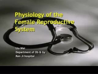
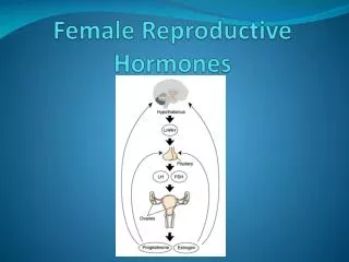
Female Reproductive Physiology. What you need to know. One axis : H-P-O axis One figure : menstrual cycle physiology. Hypothalamus. Pituitary. Ovary. Axis. Menstrual Cycle Physiology. LH. P. FSH. Hormone variation. E 2. Ovarian cycle. Uterine cycle.

E N D
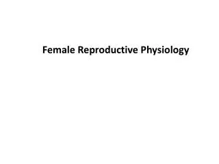
What you need to know • One axis: H-P-O axis • One figure : menstrual cycle physiology
Hypothalamus Pituitary Ovary Axis
Menstrual Cycle Physiology LH P FSH Hormone variation E2 Ovarian cycle Uterine cycle
Female development Fetal period Menopausal transition period Neonatal period Adolescencepuberty Postmenopausal period Sexual maturity childhood
Female development Fetal period Ovary develops during 8-10 week’s of pregnancy Neonatal period Within 4 weeks after birth Temporary lactation or vaginal bleeding may occur Childhood 4 weeks after birth → 10 years old Low hypothalamus - pituitary gland – ovary axis function Uterine body : cervix 1:2
Female development Adolescence / puberty 10-19 years old Onset of hypothalamus - pituitary gland – ovary axis function Uterine body : cervix 2:1 Development of second sexual characteristics Thelarche Adrenarche Growth spurt Menarche
Female development Sexual maturity From 18 years old and lasts for about 30 years Mature hypothalamus - pituitary gland – ovary axis function Reproductive age Menopausal transition period Lasts 1-10 years till menopause Declined ovarian function Vasomotor symptoms Postmenopausal period Ceased ovarian function
Hypothalamus Pituitary Thyroid Adrenal glands Ovaries
Major secretory products of the hypothalamus ----pituitary-releasing factors Gonadotropin-releasing hormone (GnRH) Luteinizing hormone (LH) Follicle-stimulating hormone (FSH) Corticotropin-releasing hormone (CRH) Adrenocorticotrophic hormone (ACTH) Growth hormone–releasing hormone (GHRH) Growth hormone (GH) Thyrotropin-releasing hormone (TRH) Thyroid-stimulating hormone (TSH)
Gonadotropin-releasing Hormone (GnRH) A decapeptide produced by hypothalamus Simultaneously regulates the secretion FSH and LH Must be secreted in a pulsatile fashion to be effective Continual exposure to GnRH results in downregulation of GnRH receptors in gonadotroph cells
Gonadotropin-releasing Hormone Extremely short half-life (only 2–4 minutes) The pulsatile secretion varies in both frequency and amplitude throughout the menstrual cycle GnRH agonist & antagonist----medical castration
Pituitary portal vessels ----bidirectional feedback control between the hypothalamus and pituitary. Anterior pituitary (adenohypophysis) Intermediate part Posterior neural pituitary (neurohypophysis)
Major secretory products of the anterior pituitary Gonadotropins: FSH,LH Growth factor (GH) Prolactin (PRL) ACTH TSH
Gonadotropins FSHFolicullar stimulating hormone LH Luteinizing hormone Responsible for ovarian follicular stimulation FSH,LH,TSHand HCG share the same a -subunit HCG
Prolactin Responsible for the synthesis of milk by the breast Stimulated by: breast manipulation, drugs, stress, exercise, and certain foods Hyperprolactinemia : amenorrhea galactorrhea Thyroid-stimulating Hormone Stimulates release of T3 and T4 from the thyroid gland Abnormalities of thyroid secretion (both hyper- and hypothyroidism) are frequently associated with ovulatory dysfunction
Adrenocorticotrophic Hormone Secreted in response to CRH Stimulates the release of adrenal glucocorticoids. Diurnal variation : early morning peak and a late evening nadir Negatively regulated by feedback from cortisol. Growth Hormone Greatest absolute amount of the anterior pituitary hormone Secreted in response to GHRH, thyroid hormone and glucocorticoids Secreted in a pulsatile fashion with peak release occurring during sleep.
Major secretory products of the posterior neural pituitary Oxytocin Arginine-vasopressin
Oxytocin A nine–amino acid peptide Stimulates of uterine muscular contraction; breast lactiferous duct myoepithelial contractions Stimulated by suckling
Arginine-vasopressin (antidiuretic hormone, or ADH, AVP) Major function : increase blood pressure arteriolar vasoconstriction renal free-water conservation decrease in blood osmolality
Menstrual Cycle Physiology LH P E2 Hormone variation FSH Ovarian cycle Uterine cycle
Menstrual cycle Normal menstrual cycle orderly cyclic hormone production parallel proliferation of the uterine lining prepare for implantation of the embryo Disorders of the menstrual cycle / menstrual physiology infertility recurrent miscarriage Irregular bleeding Malignancy
Menstrual cycle Follicular phase Luteal phase Ovarian cycle Uterine cycle Proliferative phase Secretory phase
Menstrual cycle Follicular phase development of a single dominant follicle, which should be mature at midcycle and prepared for ovulation. average length : 10 to 14 days variable in length Proliferative phase Luteal phase the time from ovulation to the onset of menses an average length of 14 days Secretory phase Normal menstrual cycle 21 to 35 days, with 2 to 6 days of flow an average blood loss of 20 to 60 mL
Hormone variation Beginning of menstrual cycle Low gonadal steroids FSH begins to rise with a cohort of growing follicles recruited Follicles secrets estrogen↑---- stimulates uterine endometrial proliferation Midpoint of the follicular phase Rising estrogen and inhibin-B inhibits pituitary FSH secretion Late in the follicular phase High estrogen stimulates LH secretion (biphasic response). Before ovulation FSH-induced LH receptors are present on granulosa cells LH stimulates progesterone secretion Estrogenic stimulation triggers pituitary LH surge, causes ovulation 24 to 36 hours later LH P E2 FSH
HormoneVariation Ovulation Heralds the transition to the luteal–secretory phase Early luteal phase Estrogen level decreases Midluteal phase Estrogen, inhibin-A increase (secreted by the corpus luteum) Progesterone levels rise precipitously after ovulation : presumptive sign of ovulation Progesterone, estrogen, and inhibin-A act centrally to suppress gonadotropin secretion and new follicular growth. remain elevated through the lifespan of the corpus luteum and then wane with its demise LH P E2 FSH
Uterine cycle Proliferative phase Secretory phase
Cyclic Changes of the Endometrium Stratum compactum decidua functionalis stratum spongiosum decidua basalis Loss of function myometrium Asherman's Syndrome
Cyclic Changes of the Endometrium Proliferative Phase progressive mitotic growth of the decidua functionalis in response to rising circulating levels of estrogen endometrial glands: straight, narrow, short →→ longer, tortuous structures mitotic cells lining proliferating glands: low columnar pattern →→ pseudostratified pattern stroma: dense compact layer vascular structures: infrequently seen
Cyclic Changes of the Endometrium Secretory Phase Ovulation occurs 14 days before mense Endometrium shift to secretory phase within 48 to 72 hours following ovulation in response to progesterone secretion Stroma: progressive increase in edema at approximately the seventh postovulatory day Spiral arteries progressively lengthen and coil Pseudodecidual d24
Secretory Phase Presence of eosinophilic protein-rich secretory products in the glandular lumen Acid–Schiff positive–staining, glycogen-containing vacuoles. Leukocytic infiltration heralds the collapse of the endometrial stroma and the onset of the menstrual flow (2 days before mense) Cyclic Changes of the Endometrium
Menses In the absence of implantation Shedding of decidua functionalis is termed menses. The destruction of the corpus luteum and its production of estrogen and progesterone is the presumed cause of the shedding. Prostaglandins release: vasospasm ; endometrial ischemia; myometrial contractions Cyclic Changes of the Endometrium
Number of oocytes during life time oogonia atresia.
Development of oocyte Enter Meiosis Ⅰ Finish Meiosis Ⅰ Secondary oocyte Primary oocyte oogonia Enter Meiosis Ⅱ birth ovulation • Meiotic arrest • 16-20 weeks of gestation • Stops at prophase I of meiosis I • Meiotic resumption • Meiosis resumes until the time of ovulation
Development of oocyte Oogonia Primary oocyte (Primordial follicle) Birth Ovulation
Follicle Theca cells: LH-R(+), produce sex steroids Granulosa cells: FSH-R,E-R,A-R,LH-R,PRL-R (+)
Follicle development Primordial follicles Preantral follicles Antral follicles preovulatoryfollicle decades 9 months Primary oocyte 3 months Follicular phase Ovulation Dominant follicle recruitment selection
Primordial follicle Preantral follicle FSH-R E-R A-R LH-R Antral follicle FSH stimulation Recruitment Selection Dominant follicle FSH-R E-R A-R LH-R PRL-R Preovulatory follicle (18-23mm) cumulus oophorus.
Two-cell Two-gonadotropin Theory there is a subdivision and compartmentalization of steroid hormone synthesis activity in the developing follicle LH cholesterol theca cells androgen FSH aromatase granulosa cells estrogen
Preovulatory Follicle Fluid-filled antrum The oocyte remains connected to the follicle by the cumulus oophorus. Rising estrogen → → negative feedback on FSH secretion Estrogen has biphasic regulation on LH Lower level → → inhibit LH secretion Sustained High level((200 pg/mL) for more than 48 hours) → → enhances LH release
Ovulation LH surge → → initiation of ovulation Occur in the single mature, or Graafian, follicle 10 to 12 hours after the LH peak or 34 to 36 hours after the initial rise in midcycle LH Dramatic increase in local concentrations of prostaglandins and proteolytic enzymes in the follicular wall Slow extrusion of the oocyte through perforation of follicular wall