Download
1 / 23
333 likes | 1.02k Views
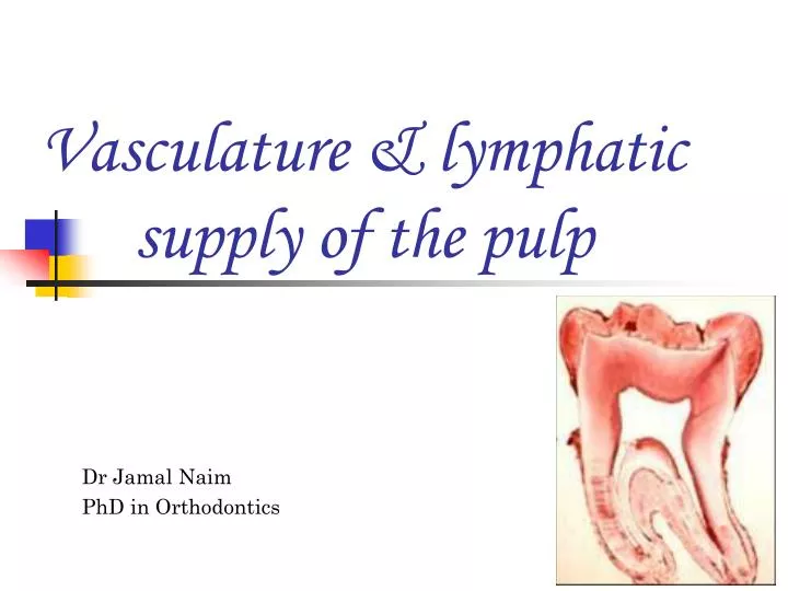
Vasculature & lymphatic supply of the pulp. Dr Jamal Naim PhD in Orthodontics. Blood supply. The pulp is highly vascularized. It is supplied by the superior and inferior alveolar artries. The blood vessels enter the pulp by the apical foramen and the accessory foramina.

E N D
Vasculature & lymphatic supply of the pulp Dr Jamal Naim PhD in Orthodontics
Blood supply • The pulp is highly vascularized. • It is supplied by the superior and inferior alveolar artries. • The blood vessels enter the pulp by the apical foramen and the accessory foramina. • One or somtimes two vessels of arterioler size (150 µm) enter the foramen accompanied with nerve bundles.
Blood supply • Blood vessels that enter the pulp through the minor accessory canals are normally not accompanied with nerve bundles. • Venules leave the pulp are associated closley with the arterioles.
Blood supply • Once the arterioles enter the pulp, an increase in the lumen and a reduction of thickness of the vessel walls occur. • The arterioles give off numerous branches along their course in the radicular pulp. • In the coronal pulp the number of branches increases, so that an intensive capillary network is formed
Blood supply • This plexus of capillaries is located at the subodontoblastic area (cell free zone of weil)
Blood supply • The capillaries adjacent to the odontoblasts are fenestrated. Those fenestration are found in areas of extensive exchange.
Blood supply • The endothelial wall of the capillaries is surrounded at random intervals with pericytes (rouget´s cells). • They are contractile cells capable of reducing the size of the vessel lumen. endothelial wall Capillary lumen Pericytes
Blood supply • Arteiovenous anastomoses (shunts) allow a direct communication between arterioles and venules. • The efferent side of circulation is comparable to those of arterioles. • The endothelial walls of of venules are much thinner with less muscle cells and comparatively larger Lumen.
Blood supply venules arterioles pulp of dog premolar
Innervation of the pulp • Several nerve bundles pass into each root via the apical foramen. • The majority of nerve fibers are unmyelinated sympathetic nerves. • They control the contraction of the smooth muscles of the vessel wall and are responsible for reductions in pulp blood flow when stimulated. • The remaining nerves are myelinated sensory nerves of the trigeminal system.
Innervation of the pulp • The myelinated nerve fibers branch extensively beneath the cell-rich zone (in the cell free zone) to form the so-called plexus of Raschkow. Plexus of Raschkow
Innervation of the pulp • In the root, no corresponding plexus exists.
Innervation of the pulp • Many myelinated fibers lose their myelin sheath and pass through the cell-free zone to terminate as receptors or as free nerve endings near odontoblasts; others pass between odontoblasts to travel a short distance up the dentinal tubules adjacent to odontoblastic processes. Plexus of Raschkow
Innervation of the pulp • Significantly, sensory nerves of the pulp respond to noxious stimuli with pain sensation only, regardless of the stimulus. • This pain is produced whether the stimulus is applied to dentin or the pulp.
Age changes in the pulp Reticular atrophy: a production of less vital pulp tissues and less response to stimulation.
Age changes in thevpulp • Continuous dentin deposition bring a decrease in the volume of the pulp cavity. • The root canal can occasionally obliterate completely. • So the blood supply is reduced, which initiate many other changes; such as: young old
Age changes in thevpulp • Reduction in cell number and activity. • Loss and degeneration of nerve axons (loss of sensitivity). • Change in the distribution of the collagen fibers. • Dystrophic calcification. young old
True denticles • True pulp stones are islands of dentin, demonstrating tubules and formative odontoblasts on their surface. • They are rare and small in size and almost located near the apical foramen. • Remnants of the epithelial root sheath invade the pulp tissue causing differentiating of the UMC to odontoblasts to form irregular dentin. odontoblasts Dentin canals
False denticles • Located more in pulp chamber • Degenerating cells or areas of hemorrhage are the nidus for calcification. • No tubules • Overdose of Vit D may cause such denticles.