Download
1 / 36
520 likes | 1.63k Views
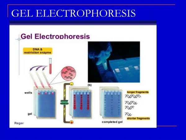
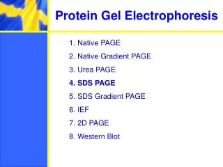
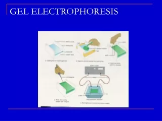
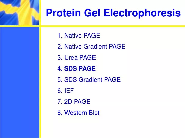
Protein Gel Electrophoresis. Native PAGE Native Gradient PAGE Urea PAGE SDS PAGE SDS Gradient PAGE IEF 2D PAGE Western Blot. Principle. Proteins move in the electric field. Their relative speed depends on the charge, size, and shape of the protein. From large to small and simple.

E N D
Protein Gel Electrophoresis • Native PAGE • Native Gradient PAGE • Urea PAGE • SDS PAGE • SDS Gradient PAGE • IEF • 2D PAGE • Western Blot
Principle Proteins move in the electric field. Their relative speed depends on the charge, size, and shape of the protein
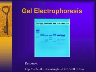
Protein visualization on gels Immediately after electrophoresis proteins in the gels are precipitated by either adding alcohol containing solutions or strong acids (e.g. TCA). Protein are often stained by Coomassie Blue dye or by photography-like treatment with AgNO3 (silver staining) There are many other stains available (e.g. Stains-all, fluorescence probes etc.)
Example of silver stained gel Silver staining is usually 10-100 times more sensitive than Coomassie Blue staining, but it is more complicated. Faint but still visible bands on this gel contain less than 0.5 ng of protein!
Native PAGE Separates folded proteins and protein-protein or protein-ligand complexes by charge, size, and shape • Useful for: • Examining protein-protein protein-ligand interactions • Detecting protein isoforms/conformers
Native PAGE examples In the absence of phospholipids, both twinfilins run as a single sharp band on this gel. PI(4,5)P2 causes twinfilin-1 and twinfilin-2 to move more rapidly toward the anode, indicating a net increase in the negative charge and thus a binding interaction. Dimerization of KIR2DL1 in the presence of Co2 Qing R. Fan et al. JBC 2000 Vartiainenet al. JBC 2003
Native gradient PAGE Separate native proteins by size – proteins stop moving when they reach a sertain gel density (but this may take a very long time ...) A great technique to study protien oligomerization!
Native gradient PAGE example Native 4-15% gradient PAGE Zavialov et al. Mol. Microbiol. 2002
Urea PAGE Separates denatured proteins by size/charge Typically 6-8 M urea is added into the gel A great technique to study protein modifications!
Example of Urea PAGE Urea PAGE of samples of heat shock protein 25 Zavialov et al. BBA1998
SDS PAGE Due to high density of binding of SDS to proteins, the ratio size/charge is nearly the same for many SDS denatured proteins. Hence proteins are separated only by length of their polypeptide chains (but not by differences in charge). Great separation. Allows estimation of the size of polypeptide chains
SDS gradient PAGE 12.5% SDS PAGE Bands in SDS gradient gel are usually sharper than in homogeneous SDS PAGE 5-20% SDS PAGE
SDS-PAGESodium Dodecyl Sulfate - Polyacrylamid Gel Electrophoresis
Sodium Dodecyl Sulfate • SDS is a common ingredient in detergents • Other names for SDS include laurel sulfate and sodium laurel sulfate • As a detergent SDS destroys protein secondary, tertiary and quaternary structure • This makes proteins rod shaped • SDS also sticks to proteins in a ratio of approximately 1.4 g of SDS for each gram of protein • Negative charge on the sulfate groups of SDS mask any charge on the protein
O H-C-C-C-C-C-C-C-C-C-C-C-C-O-S-O-Na+ O Non-polar Hydrophobic tail Polar Hydrophilic head H H H H H H H H H H H H H H H H H H H H H H H H SDSSodium Dodecyl Sulfate C12H25NaO4S • Because it is amphipathic, SDS is a potent detergent
SDS Protein SDS and Proteins
SDS and Proteins • SDS nonpolar chains arrange themselves on proteins and destroy secondary tertiary and quarternary structrure • Thus shape is no longer an issue as the protein SDS complex becomes rod shaped • In aqueous solutions, SDS polarizes releasing Na+ and retaining a negative charge on the sulfate head • So much SDS binds to proteins that the negative charge on the SDS drowns out any net charge on protein side chains • In the presence of SDS all proteins have uniform shape and charge per unit length
O Acrylamide C NH2 CH2 CH CH2 CH O C NH2 CH2 Acrylamide Acrylamide O C NH2 bis-Acrylamide CH2 CH Polyacrylamide Gels • Polyacrilamide is a polymer made of acrylamide (C3H5NO) and bis-acrilamide (N,N’-methylene-bis-acrylamide C7H10N2O2)
O O C C NH2 NH2 CH2 CH2 CH CH SO4-. Polyacrylamide Gels • Acrylamide polymerizes in the presence of free radicals typically supplied by ammonium persulfate
O O O O C NH2 C NH2 C NH2 C NH2 CH2 CH CH2 CH CH2 CH CH2 CH SO4-. Polyacrylamide Gels 1. Acrylamide polymerizes in the presence of free radicals typically supplied by ammonium persulfate • 2.TMED (N,N,N’,N’-tetramethylethylenediamine) serves as a catalyst in the reaction
O O O C NH2 C NH2 C NH2 CH2 CH CH2 CH CH2 CH CH2 CH O O O C NH2 C NH2 C NH2 CH2 O CH2 CH CH2 CH O C NH2 C NH2 CH2 CH CH2 CH bis-Acrylamide Polyacrylamide Gels • bis-Acrylamide polymerizes along with acrylamide forming cross-links between acrylamide chains
Polyacrylamide Gels • bis-Acrylamide polymerizes along with acrylamide forming cross-links between acrylamide chains
Lots of bis-acrylamide Little bis-acrylamide Polyacrylamide Gels • Pore size in gels can be varied by varying the ratio of acrylamide to bis-acrylamide • Protein separations typically use a 29:1 or 37.5:1 acrylamide to bis ratio
3 Addition of SDS 2 Protein becomes rod-shaped with uniform charge distribution 1 SDS-PAGE 1 2 3
IEF Separates proteins by their isoelectric points (pI) Each protein has own pI = pH at which the protein has equal amount of positive and negative charges (the net charge is zero)
IEF Mixtures of ampholytes, small amphoteric molecules with high buffering capacity near their pI, are used to generate the pH gradient. Positively and negatively charged proteins move to – and +, respectively, until they reach pI. PI of proteins can be theoretically predicted. Therefore, IEF can also be used for protein identification.
IEF example IEF 4-6.5 pH gradient Zavialov A.
2D PAGE Lung V79 cells from chinese hamster Guillermo Senisterra Dept. of Physics-University of Waterloo-Waterloo-Ontario N2L 3G1-Canada
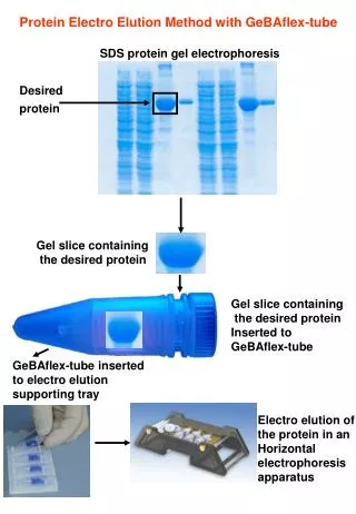
Western Blotting (WB) WB is a protein detection technique that combines the separation power of SDS PAGE together with high recognition specificity of antibodies An antibody against the target protein could be purified from serum of animals (mice, rabbits, goats) immunized with this protein Alternatively, if protein contains a commonly used tag or epitope, an antibody against the tag/epitope could be purchase from a commercial source (e.g. anti-6 His antibody)
WB: 4 steps • Separation of proteins using SDS PAGE • 2. Transfer of the proteins onto e.g. a nitrocellulose membrane (blotting) • 3. Immune reactions • 4. Visualization