Download
1 / 36
380 likes | 790 Views
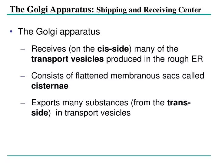
The Golgi Apparatus: Shipping and Receiving Center. The Golgi apparatus Receives (on the cis-side ) many of the transport vesicles produced in the rough ER Consists of flattened membranous sacs called cisternae Exports many substances (from the trans-side ) in transport vesicles.

E N D
The Golgi Apparatus: Shipping and Receiving Center • The Golgi apparatus • Receives (on the cis-side) many of the transport vesicles produced in the rough ER • Consists of flattened membranous sacs called cisternae • Exports many substances (from the trans-side) in transport vesicles
cis face (“receiving” side of Golgi apparatus) 5 3 4 6 2 1 Vesicles coalesce to form new cis Golgi cisternae Vesicles move from ER to Golgi 0.1 0 µm Vesicles also transport certain proteins back to ER Cisternae Cisternal maturation: Golgi cisternae move in a cis- to-trans direction Vesicles form and leave Golgi, carrying specific proteins to other locations or to the plasma mem- brane for secretion trans face (“shipping” side of Golgi apparatus) Vesicles transport specific proteins backward to newer Golgi cisternae Functions of the Golgi apparatus • Modification of the products of the rough ER • Manufacture of certain macromolecules • Probably evolved from ER Golgi apparatus Figure 6.13 TEM of Golgi apparatus
1 µm Nucleus Lysosome Hydrolytic enzymes digest food particles Food vacuole fuses with lysosome Lysosome contains active hydrolytic enzymes Digestive enzymes Lysosome Plasma membrane Digestion Food vacuole (a) Phagocytosis: lysosome digesting food Lysosomes: Digestive Compartments • Lysosomes are membranous sacs of hydrolytic enzymes, and they carry out intracellular digestion. • They digest food from food vacuoles that form by phagocytosis and they recycle old cell parts in autophagy. Figure 6.14 A
Lysosome containing two damaged organelles 1 µ m Mitochondrion fragment Peroxisome fragment Lysosome fuses with vesicle containing damaged organelle Hydrolytic enzymes digest organelle components Lysosome Digestion Vesicle containing damaged mitochondrion (b) Autophagy: lysosome breaking down damaged organelle Lysosomes • different lysosomes have different enzymes for breaking down different macromolecules • They have a low pH (around 5); pump H+ ions in from the cell • Example of a lysosomal disease: Tay-Sachs disease, caused by a missing lysosomal enzyme for lipid breakdown, leads to buildup of lipids in the brain, killing the individual in infancy. Figure 6.14 B
Vacuoles: Diverse Maintenance Compartments • Vacuoles are fluid filled and membrane enclosed. • A cell may have one or several vacuoles. • Food vacuoles • Are formed by phagocytosis • Contractile vacuoles • Pump excess water out of protist cells
Central vacuole Cytosol Tonoplast Nucleus Central vacuole Cell wall Chloroplast 5 µm Vacuoles: Diverse Maintenance Compartments • Central vacuoles • Found in plant cells • Function in cell size and turgidity • Store reserves of important organic compounds and water Figure 6.15
1 Nuclear envelope is connected to rough ER, which is also continuous with smooth ER Nucleus Rough ER 2 Membranes and proteins produced by the ER flow in the form of transport vesicles to the Golgi Smooth ER cis Golgi Nuclear envelop 3 Golgi pinches off transport Vesicles and other vesicles that give rise to lysosomes and Vacuoles Plasma membrane trans Golgi 4 5 6 Lysosome available for fusion with another vesicle for digestion Transport vesicle carries proteins to plasma membrane for secretion Plasma membrane expands by fusion of vesicles; proteins are secreted from cell The Endomembrane System: A Review • Relationships among membranes/organelles of the endomembrane system Figure 6.16
Organelles of Endosymbiotic Origin • Mitochondria and chloroplasts change energy from one form to another • Mitochondria • Are sites of cellular respiration • Plastids • Found only in plants, are sites of photosynthesis
Mitochondrion Intermembrane space Outer membrane Free ribosomes in the mitochondrial matrix Inner membrane Cristae Matrix Mitochondrial DNA 100 µm Mitochondria: Chemical Energy Conversion • Mitochondria (powerhouse of the cell) • Are found in nearly all eukaryotic cells • Have their own DNA- derived from the mother. This DNA changes very slowly over time because there is no recombination, only change is due to drift (chance). Figure 6.17
Mitochondrion Intermembrane space Outer membrane Free ribosomes in the mitochondrial matrix Inner membrane Cristae Matrix Mitochondrial DNA 100 µm Mitochondria: Chemical Energy Conversion • Are the site of oxidative metabolism (conversion of glucose to ATP, carbon dioxide, and water), also known as cellular respiration. * Which type of cell would you expect to have a lot of mitochondria? • Are enclosed in a double membrane. Inner membrane is folded for increased surface area. This is where the metabolism occurs; enzymes are embedded in the membrane. Figure 6.17
Plastids: Capture of Light Energy • Plastids • have a double membrane • have their own DNA • function in photosynthesis (the chloroplast is an example) • contain pigments such as chlorophyll, carotenoids • can also be for storage (leukoplasts)
Chloroplast Ribosomes Stroma Chloroplast DNA Inner and outer membranes Granum 1 µm Thylakoid Chloroplasts • Are found in leaves and other green organs of plants and in algae • Their structure includes • Thylakoids, membranous sacs • Stroma, the internal fluid Figure 6.18
Chloroplast Peroxisome Mitochondrion 1 µm Peroxisomes: Oxidation • Peroxisomes • Produce hydrogen peroxide and convert it to water Figure 6.19
The Cytoskeleton Cytoplasm – includes all the space inside the plasma membrane but outside the nucleus (includes organelles, cytosol, and cytoskeleton) Cytoskeleton: microlattice of fibers supports the cell and gives it 3-dimensional shape. Organelles attach to the fibers. The cytoskeleton gives the cell spatial information, which is very important in development The cytoskeleton is not stationary, it is dynamic.
Microtubule Microfilaments 0.25 µm Figure 6.20 The Cytoskeleton • Is a network of fibers extending throughout the cytoplasm, and it organizes cell structures and activities. Figure 6.20
Vesicle ATP Receptor for motor protein Motor protein (ATP powered) Microtubule of cytoskeleton (a) Motor proteins that attach to receptors on organelles can “walk” the organelles along microtubules or, in some cases, microfilaments. Vesicles Microtubule 0.25 µm (b) Vesicles containing neurotransmitters migrate to the tips of nerve cell axons via the mechanism in (a). In this SEM of a squid giant axon, two vesicles can be seen moving along a microtubule. (A separate part of the experiment provided the evidence that they were in fact moving.) Figure 6.21 A, B Roles of the Cytoskeleton: Support, Motility, and Regulation • Gives mechanical support to the cell • Is involved in cell motility, which utilizes motor proteins
Table 6.1 Components of the Cytoskeleton • There are three main types of fibers that make up the cytoskeleton
Microtubules • Microtubules • Shape the cell • Cilia and flagella for motility • Guide the movement of organelles • Help separate the chromosome copies in dividing cells
Centrosomes and Centrioles • The centrosome • Is considered to be a “microtubule-organizing center” and it organizes the spindle fibers used to guide the movement of chromosomes during cell division.
Centrosome Microtubule Centrioles 0.25 µm Longitudinal section of one centriole Cross section of the other centriole Microtubules Figure 6.22 In animal cells, the centrosome: • Contains a pair of centrioles which are made of microtubules in a nine-triplets pattern.
Outer microtubule doublet Plasma membrane 0.1 µm Dynein arms Central microtubule Outer doublets cross-linking proteins inside Microtubules Radial spoke Plasma membrane Basal body (b) 0.5 µm 0.1 µm (a) Triplet (c) Figure 6.24 A-C Cross section of basal body Cilia and flagella – locomotory organelles • Cilia and flagella share a common ultrastructure of microtubules in a 9 + 2 arrangement. The base structure is similar to that of centrioles (nine triplets).
Microtubule doublets ATP Dynein arm Powered by ATP, the dynein arms of one microtubule doublet grip the adjacent doublet, push it up, release, and then grip again. If the two microtubule doublets were not attached, they would slide relative to each other. (a) Figure 6.25 A Cilia and Flagella move through the action of motor proteins • The protein dynein • Is responsible for the bending movement of cilia and flagella
ATP Outer doublets cross-linking proteins Anchorage in cell (b) In a cilium or flagellum, two adjacent doublets cannot slide far because they are physically restrained by proteins, so they bend. (Only two of the nine outer doublets in Figure 6.24b are shown here.) Ciliary/flagellar motion Figure 6.25 B
Microvillus Plasma membrane Microfilaments (actin filaments) Intermediate filaments 0.25 µm Figure 6.26 Microfilaments (Actin Filaments) • Are built from molecules of the protein actin • Are found in microvilli
Muscle cell Actin filament Myosin filament Myosin arm (a) Figure 6.27 A Myosin motors in muscle cell contraction. Microfilaments of muscle • Microfilaments that function in cellular motility • Contain the protein myosin in addition to actin
Cortex (outer cytoplasm): gel with actin network Inner cytoplasm: sol with actin subunits Extending pseudopodium (b) Amoeboid movement Amoeboid motion • Involves the contraction of actin and myosin filaments Figure 6.27 B
Nonmoving cytoplasm (gel) Chloroplast Streaming cytoplasm (sol) Parallel actin filaments Cell wall (b) Cytoplasmic streaming in plant cells Cytoplasmic streaming • Is another form of locomotion created by microfilaments Figure 6.27 C
Intermediate Filaments • Support cell shape • Fix organelles in place • Are fixed and do not disassemble.
Extracellular components and connectionsbetween cells help coordinate cellular activities
Central vacuole of cell Plasma membrane Secondary cell wall Primary cell wall Central vacuole of cell Middle lamella 1 µm Central vacuole Cytosol Plasma membrane Plant cell walls Plasmodesmata Figure 6.28 Cell Walls of Plants • Extracellular structures of plant cells that distinguish them from animal cells • Are made of cellulose fibers embedded in other polysaccharides and protein • May have multiple layers
The Extracellular Matrix (ECM) of Animal Cells • Animal cells • Lack cell walls • Are covered by an elaborate matrix, the ECM
Polysaccharide molecule EXTRACELLULAR FLUID Collagen A proteoglycan complex Carbo- hydrates Core protein Fibronectin Proteoglycan molecule Plasma membrane Integrins CYTOPLASM Micro- filaments Integrin Figure 6.29 The ECM • Is made up of glycoproteins and other macromolecules. Some of these molecules can be part of self-recognition or membrane-membrane interactions (e.g. tissue glue that holds cells together).
Functions of the ECM include • Support • Adhesion • Movement • Regulation
Cell walls Interior of cell Interior of cell 0.5 µm Plasmodesmata Plasma membranes Figure 6.30 Intercellular Junctions in Plants • Plasmodesmata are channels that perforate plant cell walls. The cell membranes of neighboring cells are continuous through these pores in the cell walls. This allows cells to share molecules and communicate.
Animal Cell Junctions • In animals, there are three types of intercellular junctions • Tight junctions • Desmosomes • Gap junctions
TIGHT JUNCTIONS At tight junctions, the membranes of neighboring cells are very tightly pressed against each other, bound together by specific proteins (purple). Forming continu- ous seals around the cells, tight junctions prevent leakage of extracellular fluid across A layer of epithelial cells. Tight junction Tight junctions prevent fluid from moving across a layer of cells 0.5 µm DESMOSOMES Desmosomes (also called anchoring junctions) function like rivets, fastening cells Together into strong sheets. Intermediate Filaments made of sturdy keratin proteins Anchor desmosomes in the cytoplasm. Tight junctions Intermediate filaments Desmosome Gap junctions 1 µm GAP JUNCTIONS Gap junctions (also called communicating junctions) provide cytoplasmic channels from one cell to an adjacent cell. Gap junctions consist of special membrane proteins that surround a pore through which ions, sugars, amino acids, and other small molecules may pass rapidly. Gap junctions are necessary for commu- nication between cells in many types of tissues, including heart muscle and animal embryos. Extracellular matrix Space between cells Gap junction Plasma membranes of adjacent cells Figure 6.31 0.1 µm Animal Cell Junctions • Types of intercellular junctions in animals