Download
1 / 35
420 likes | 481 Views
Explore the general functions and anatomy of the urinary system, focusing on the nephron's working mechanism and control. Learn about micturition processes, urine production, fluid regulation, and hormone production in the kidneys.

E N D
Kidney Physiology Fabian Omenankiti
Lecture Outline • General Functions of the Urinary System • Quick overview of the functional anatomy of the urinary system • How the nephron works & is controlled • Micturition
General Functions • Produce & expel urine • Regulate the volume and composition of the extracellular fluid • Control pH • Control blood volume & blood pressure • Controls osmolarity • Controls ion balance • Production of hormones • Renin • EPO
Overview of Function AnatomyThe System • Urinary system consists of: Kidneys – The functional unit of the system Ureters Conducting & Storage components Urinary Bladder Urethra
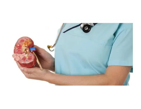
Overview of Functional AnatomyThe Kidney • Divided into an outer cortex • And an inner medulla • The functional unit of this kidney is the nephron • Which is located in both the cortex and medullary areas renal pelvis
Overview of Functional AnatomyThe Kidney • The nephron consists of: • Vascular components • Afferent & efferent arterioles • Glomerulus • Peritubular capillaries • Vasa recta • Tubular components • Proximal convoluted tubule • Distal convoluted tubule • Nephron loop (loop of Henle) • Collecting duct • Tubovascular component • Juxtaglomerular appartus
The Nephron • Simplified view of its functions • Glomerular Filtration • Tubular Reabsorption • Tubular Secretion • Excretion
The Nephron • Locations for filtration, reabsorption, secretion & excretion
NephronFiltration • First step in urine formation • No other urinary function would occur without this aspect! • Occurs in the glomerulus due to • Filtration membrane & • Capillary hydrostatic pressure • Colloid osmotic pressure • Capsular hydrostatic pressure
NephronFiltration Membrane • Capillaries are fenestrated • Overlying podocytes with pedicels form filtration slits • Basement membrane between the two
NephronGlomerular Filtration • Barriers • Mesanglial cells can alter blood flow through capillaries • Basal lamina alters filtration as well by • Containing negatively charged glycoproteins • Act to repel negatively charged plasma proteins • Podocytes form the final barrier to filtration by forming “filtration slits”
NephronGlomerular Filtration • Forces • Blood hydrostatic pressure (PH) • Outward filtration pressure of 55 mm Hg • Constant across capillaries due to restricted outflow (efferent arteriole is smaller in diameter than the afferent arteriole) • Colloid osmotic pressure (π) • Opposes hydrostatic pressure at 30 mm Hg • Due to presence of proteins in plasma, but not in glomerular capsule (Bowman’s capsule) • Capsular hydrostatic pressure (Pfluid) • Opposes hydrostatic pressure at 15 mm Hg
NephronGlomerular Filtration • 10 mm Hg of filtration pressure • Not high, but has a large surface area and nature of filtration membrane • creates a glomerular filtration rate (GFR) of 125 ml/min which equates to a fluid volume of 180L/day entering the glomerular capsule. • Plasma volume is filtered 60 times/day or 2 ½ times per hour • Requires that most of the filtrate must be reabsorbed, or we would be out of plasma in 24 minutes! • Still…. GFR must be under regulation to meet the demands of the body.
NephronGlomerular Filtration • 10 mm Hg of filtration pressure • Not high, but has a large surface area and nature of filtration membrane • creates a glomerular filtration rate (GFR) of 125 ml/min which equates to a fluid volume of 180L/day entering the glomerular capsule. • Plasma volume is filtered 60 times/day or 2 ½ times per hour • Requires that most of the filtrate must be reabsorbed, or we would be out of plasma in 24 minutes! • GFR maintains itself at the relatively stable rate of 180L/dayby • Regulation of blood flowthrough the arterioles • Changing afferent andefferent arterioles hasdifferent effects on GFR
NephronRegulation of GFR • How does GFR remain relatively constant despite changing mean arterial pressure? 1. Myogenic response • Typical response to stretch of arteriolar smooth muscle due to increased blood pressure: • increase stretch results in smooth muscle contraction and decreased arteriole diameter • Causes a reduction in GFR • If arteriole blood pressure decreases slightly, GFR only increases slightly as arterioles dilate • Due to the fact that the arterioles are normally close to maximal dilation • Further drop in bp (below 80mmHg) reduced GFR and conserves plasma volume 2. Tubulooglomerular feedback at the JGA 3. Hormones & ANS
NephronAutoregulation of GFR 2. Tubulooglomerular feedback at the JGA • Fluid flow is monitored in the tubule where it comes back between the afferent and efferent arterioles • Forms the juxtaglomerular apparatus • Specialized tubular cells in the JGA form the macula densa • Specialized contractile cells in the afferent arteriole in the JGA are called granular cells or juxtaglomerular cells
NephronRegulation of GFR • The cells of the macula densa monitor NaCl concentration in the fluid moving into the dital convoluted tubule. • If GFR increases, then NaCl movement also increases as a result • Macula densa cells send a paracrine message (unknown for certain) causing the afferent arteriole to contract, decreasing GFR and NaCl movment
NephronRegulation of GFR • Hormones & ANS • Autoregulation does a pretty good job, however extrinsic control systems can affect a change by overriding local autoregulation factors by • Changing arteriole resistance • Sympathetic innervation to both afferent and efferent arterioles • Acts on alpha receptors causing vasoconstriction • Used when bp drops drastically to reduce GFR and conserve fluid volume • Changing the filtration coefficient • Release of renin from the granular cells (JG cells) of the JGA initiates the renin-angiotensin-aldosterone system (RAAS) • Angiotensin II is a strong vasoconstrictor • Prostaglandins • Vasodilators • These hormones may also change the configuration of the mesanglial cells and the podocytes, altering the filtration coefficient
NephronRegulation of GFR • Renin-Angiotensin-Aldosterone System (RAAS) (or low NaCl flow in JGA)
NephronTubular Reabsorption • GFR = 180 L/day, >99% is reabsorbed • Why so high on both ends? • Allows material to be cleared from plasma quickly and effectively if needed • Allows for easy tuning of ion and water balance • Reabsorption • Passive and Active Transport Processes • Most of the reabsorption takes place in the PCT Movement may be via epithelial transport (through the cells) or by paracellular pathways (between the epithelial cells)
NephronTubular Reabsorption • Na+ reabsorption • An active process • Occurs on the basolateral membrane (Na+/K+ ATPase) • Na+ is pumped into the interstitial fluid • K+ is pumped into the tubular cell • Creates a Na+ gradient that can be utilized for 2º active transport
NephronTubular Reabsorption • Secondary Active Transport utilizing Na+ gradient (Sodium Symport) • Used for transporting • Glucose, amino acids, ions, metabolites
NephronTubular Reabsorption • The transport membrane proteins • Will reach a saturation point • They have a maximum transport rate = transport maximum (Tm) • The maximum numberof molecules that can betransported per unit oftime • Related to the plasmaconcentration called therenal threshold… • The point at whichsaturation occurs andTm is exceeded
NephronTubular Reabsorption • Glucose Reabsorption • Glucose is filtered and reabsorbed hopefully 100% • Glucose excreted = glucose filtered – glucose reabsorbed Implication of no glucose transports past the PCT?
NephronTubular Reabsorption • Where does filtered material go? • Into peritubular capillaries because in the capillaries there exists • Low hydrostatic pressure • Higher colloid osmotic pressure
NephronTubular Secretion • Tubular secretion is the movement of material from the peritubular capillaries and interstitial space into the nephron tubules • Depends mainly on transport systems • Enables further removal of unwanted substances • Occurs mostly by secondary active transport • If something is filtered, not reabsorbed, and secreted… the clearance rate from plasma is greater than GFR! • Ex. penicillin – filtered and secreted, not reabsorbed • 80% of penicillin is gone within 4 hours after administration
NephronExcretion & Clearance Filtration – reabsorption + secretion = Excretion • The excretion rate then of a substance (x) depends on • the filtration rate of x • if x is reabsorbed, secreted or both • This just tells us excretion, but not much about how the nephron is working in someone • This is done by testing a known substance that should be filtered, but neither reabsorbed or secreted • 100% of the filtered substance is excreted and by monitoring plasma levels of the substance, a clearance rate can be determined
NephronExcretion & Clearance • Inulin • A plant product that is filtered but not reabsorbed or secreted • Used to determine clearance rate and therefore nephron function
NephronExcretion & Clearance • The relationship between clearance and excretion using a few examples
NephronUrine Concentration & Dilution • Urine normally exits the nephron in a dilute state, however under hormonal controls, water reabsorption occurs and can create an extremely concentrated urine. • Aldosterone & ADH are the two main hormones that drive this water reabsorption • Aldosterone creates an obligatory response • Aldosterone increases Na+/K+ ATPase activity and therefore reabsorption of Na+… where Na+ goes, water is obliged to follow • ADH creates a facultative response • Opens up water channels in the collecting duct, allowing for the reabsorption of water via osmosis
Micturition • Once excreted, urine travels via the paired ureters to the urinary bladder where it is held (about ½ L) • Sphincters control movement out of the bladder • Internal sphincter – smooth muscle (invol.) • External sphincter – skeletal muscle (vol.)
Micturition • Reflex Pathway