Download

1 / 35
350 likes | 619 Views
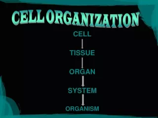
Cell Theory. Cells are basic units of organization and function in all living organisms All cells come from other cellsAll living cells have evolved from a common ancestor . Homeostasis. Cells have many organelles, internal structures that carry out specific functions help maintain homeostasis.

E N D
1. Organization of the Cell Chapter 4 2nd edit BTT2 3/06 AD2nd edit BTT2 3/06 AD
2. Cell Theory Cells are basic units of organization and function in all living organisms
All cells come from other cells
All living cells have evolved from a common ancestor
3. Homeostasis
Cells have many organelles,
internal structures that carry out specific functions
help maintain homeostasis
4. Plasma Membrane Plasma membrane
surrounds the cell
separates cell from external environment
maintains internal conditions
allows the cell to exchange materials with outer environment
5. Prokaryotes Prokaryotic cells
No internal membrane organization
nuclear area (not nucleus)
cell wall
ribosomes
flagella
6. Figure 4.6: Structure of a prokaryotic cell.
This colorized TEM shows a thin lengthwise slice through an Escherichia coli bacterium. Note the prominent nuclear area containing the genetic material (DNA). E. coli is a normal inhabitant of the human intestine, but under certain conditions some strains can cause infections.Figure 4.6: Structure of a prokaryotic cell.
This colorized TEM shows a thin lengthwise slice through an Escherichia coli bacterium. Note the prominent nuclear area containing the genetic material (DNA). E. coli is a normal inhabitant of the human intestine, but under certain conditions some strains can cause infections.
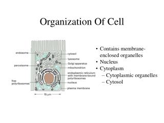
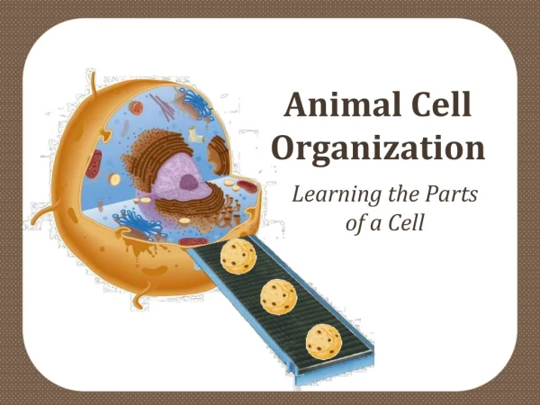
7. Eukaryotes Eukaryotic cells
membrane-enclosed nucleus
cytoplasm contains organelles
cytosol (fluid component)
8. Figure 4.8: Composite diagram of an animal cell.
This generalized animal cell is shown in a realistic context surrounded by adjacent cells, which cause it to be slightly compressed. The TEMs show the structure of various organelles. Depending on the cell type, certain organelles may be more or less prominent.Figure 4.8: Composite diagram of an animal cell.
This generalized animal cell is shown in a realistic context surrounded by adjacent cells, which cause it to be slightly compressed. The TEMs show the structure of various organelles. Depending on the cell type, certain organelles may be more or less prominent.
9. Plant Cells Plant cells
rigid cell walls
large vacuoles
plastids
chloroplasts
mitochondria
10. Figure 4.7: Composite diagram of a plant cell.
Chloroplasts, a cell wall, and prominent vacuoles are characteristic of plant cells. The TEMs show specific structures or areas of the cell. Some plant cells do not have all the organelles shown here. For example, leaf and stem cells that carry on photosynthesis contain chloroplasts, whereas root cells do not. Many of the organelles, such as the nucleus, mitochondria, and endoplasmic reticulum (ER), are characteristic of all eukaryotic cells.Figure 4.7: Composite diagram of a plant cell.
Chloroplasts, a cell wall, and prominent vacuoles are characteristic of plant cells. The TEMs show specific structures or areas of the cell. Some plant cells do not have all the organelles shown here. For example, leaf and stem cells that carry on photosynthesis contain chloroplasts, whereas root cells do not. Many of the organelles, such as the nucleus, mitochondria, and endoplasmic reticulum (ER), are characteristic of all eukaryotic cells.
11. Cell Membranes Divide cell into compartments
Vesicles:
transport materials between compartments
Endomembrane system
Rough ER
Smooth ER
Golgi apparatus
12. The Nucleus Control center of cell
genetic information coded in DNA
Nuclear envelope
double membrane
Nuclear pores
communicate with cytoplasm
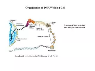
13. Nuclear Structures Chromatin
DNA and protein
Chromosomes
DNA condensed for cell division
Nucleolus
ribosomal RNA synthesis
ribosome assembly
14. The Nucleus
15. Endoplasmic Reticulum (ER) Network of folded membranes
in cytosol
Smooth ER
lipid synthesis
calcium ion storage
detoxifying enzymes
Rough ER
ribosomes on outer surface
produces proteins
16. ER
17. The Golgi Complex Processes proteins synthesized by ER
Manufactures lysosomes
Cisternae
stacks of flattened membranous sacs
18. Other Organelles Lysosomes
enzymes break down structures
Vacuoles
store materials in plant cells
Peroxisomes
produce and degrade hydrogen peroxide (catalase)
19. Mitochondria Site of aerobic respiration
Double membrane
inner membrane folded (cristae)
matrix (cristae and inner compartment)
20. Mitochondria
21. Plastids Plastids
organelles that produce and store food
in cells of plants and algae
Chloroplasts
plastids that carry out photosynthesis
22. Chloroplast Structure Stroma
fluid-filled space enclosed by inner membrane of chloroplast
Grana
stacks of membranous sacs (thylakoids)
suspended in stroma
23. Chloroplasts
24. Photosynthesis Chlorophyll
green pigment in thylakoid membranes
traps light energy
Light energy converted to chemical energy in ATP
used to synthesize carbohydrates from carbon dioxide and water
25. The Cytoskeleton Microtubules
hollow tubulin cylinders
role in cell division
Microfilaments
actin filaments
important in cell movement
Intermediate filaments
strengthen cytoskeleton
stabilize cell shape
26. Cytoskeleton
27. Centrosome Anchor microtubules
Usually contains two centrioles
Each centriole has 9 x 3 arrangement of microtubules
28. Figure 4.24: Centrioles.Figure 4.24: Centrioles.
29. Cilia and Flagella Cilia and flagella
thin, movable structures
project from cell surface
function in movement
Cilia are short�flagella are long
30. Flagella Bacterial flagella:
composed of flagellin
rotates like a propeller
Eukaryotic flagella:
composed of microtubules
9 + 2 arrangement
wave back and forth
31. Figure 4.25: Structure of cilia.Figure 4.25: Structure of cilia.
32. Cell Coat Glycocalyx (cell coat)
Surrounds cell
Polysaccharides extend from plasma membrane
33. ECM Extracellular matrix (ECM)
Surrounds many animal cells
Carbohydrates and protein
Fibronectins
glycoproteins of ECM
bind to integrins
Integrins
receptor proteins in plasma membrane
34. ECM
35. Cell Wall Cellulose & other polysaccharides
form rigid cell walls
in bacteria, fungi, and plant cells
36. Figure 4.29: Plant cell walls.
The cell walls of two adjacent plant cells are labeled in this TEM. The cells are cemented together by the middle lamella, a layer of gluelike polysaccharides called pectins. A growing plant cell first secretes a thin primary wall that is flexible and can stretch as the cell grows. The thicker layers of the secondary wall are secreted inside the primary wall after the cell stops elongating.Figure 4.29: Plant cell walls.
The cell walls of two adjacent plant cells are labeled in this TEM. The cells are cemented together by the middle lamella, a layer of gluelike polysaccharides called pectins. A growing plant cell first secretes a thin primary wall that is flexible and can stretch as the cell grows. The thicker layers of the secondary wall are secreted inside the primary wall after the cell stops elongating.