Download
1 / 98
1.05k likes | 1.75k Views
Pericardial Disease. Two-Dimensional Echocardiographic and Doppler Findings. Pericardial Disease. Pericardium Fibrous Pericardium Serous Pericardium - Parietal Pericardium - Visceral Pericardium. Pericardial Disease. Pericardial Sac (Space)

E N D
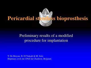
Pericardial Disease Two-Dimensional Echocardiographic and Doppler Findings
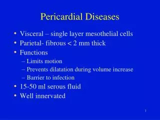
Pericardial Disease Pericardium • Fibrous Pericardium • Serous Pericardium - Parietal Pericardium - Visceral Pericardium
Pericardial Disease Pericardial Sac (Space) • Space between parietal and visceral pericardium. • Around the RV and LV – ellipsoid structure conforming to the ventricular apex.
Pericardial Disease Pericardial Sac (Space) • Around the pulmonary and systemic venous inflows and around the great vessels – the pericardia meet to close the end of the sac – pericardial reflections
Pericardial Disease Pericardial Sinuses • Transverse sinus • Oblique sinus
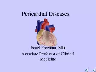
Pericarditis Inflammation of the pericardium.
Pericarditis Causes • Infection – viral and bacteria • Trauma • Uremia • Transmural infarct
Pericarditis Diagnosis • Chest pain • EKG changes • Presence of pericardial friction rub on auscultation.
Pericarditis Pericardial effusion is not a necessary criterion for the diagnosis of pericarditis.
Pericardial Effusion Infection • Postviral pericarditis • Bacterial • Tuberculosis
Pericardial Effusion Malignant • Metastatic disease – lymphoma & melanoma • Direct extension – lung and breast • Primary cardiac malignancy
Pericardial Effusion Inflammatory • Post MI (Dressler’s Syndrome) • Uremia • Collagen vascular disease • Post cardiac surgery
Pericardial Effusion Intracardiac-Pericardial Communication • Blunt or penetrating chest trauma • Post catheter procedures • LV rupture (post MI)
Pericarditis Echocardiographic Findings • Pericardial effusion • Pericardial thickening +/- effusion • Normal
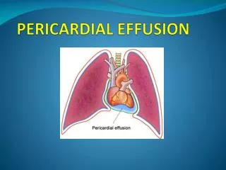
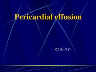
Pericarditis Pericardial Effusion Seen as an echolucent space around the heart.
Pericarditis Pericardial thickening • Increased echogenicity of the pericardial reflection on 2D imaging. • Multiple parallel reflections posterior to the LV on the M-mode recordings.
Pericarditis • Pericarditis is a clinical diagnosis that cannot be made independently by echocardiagraphy. • Goal of echocardiography is to evaluate for pericardial effusion or thickening.
Pericardial Tamponade Tamponade Physiology • Occurs when the pressure in the pericardium exceeds the pressure in the cardiac chambers, resulting in impair cardiac filling.
Pericardial Tamponade Physiology • The physiologic consequences of fluid in the pericardial space depends on: - the volume - the rate of accumulation.
Pericardial Tamponade • As pericardial pressure increases, filling of each cardiac chamber is sequentially impaired, with the lower-pressure chamber (atria) affected before the higher-pressure chambers.
Pericardial Disease The compressive effect of the pericardial fluid is seen most clearly in the phase of the cardiac cycle when pressure is lowest in that chamber. - systole for the atria - diastole for the ventricles
Pericardial Disease In response to the increase pericardial pressure: The filling pressures become elevated as a compensatory mechanism to maintain cardiac output.
Pressure Changes In fully developed tamponade All four cardiac chambers are equal and elevated due to exposure of the entire heart to the elevated pericardial pressure.
Clinical Findings Clinical Findings • Low cardiac output • Hypotension
Clinical Findings • Tachycardia • Elevated JVP • Pulsus paradoxus (inspiratory decline > 10 mmHg in systemic blood pressure)
2-D Findings • Pericardial effusion – moderate to large. • RA systolic collapse (duration > 1/3 systole). • RV diastolic collapse.
2-D Findings • Reciprocal changes in right and left ventricular volume with respiration. • Varying pattern of septal motion. • IVC plethora.
Doppler Findings Respiratory variation in right and left diastolic filling: • Increased RV filling on the first beat after inspiration. • Decreased LV filling on the first beat after inspiration.
Doppler Findings • Increase flow velocity integral of the PA in inspiration. • Decrease flow velocity integral of the aorta in inspiration.
Pericardial Effusion Effusion • Echolucent space adjacent to cardiac structure. • Diffuse and symmetrical (in the absence of prior pericardial disease or surgery).
Pericardial Effusion A relative echogenic area anteriorly, in the absence of a posterior effusion most likely represents a pericardial fat pad.
Pericardial Effusion M-mode findings • Flat posterior pericardial echo reflection. • Moving epicardial echo. • Separation between the two both in systole and diastole.
Pericardial Effusion Fibrinous Stranding • Recurrent or longstanding pericardial disease. • Metastatic disease pericardial disease.
Pericardial Effusion Metastatic disease • Fibrinous strand formation. • Nodular appearance, evidence of extension into the myocardium. • Appropriate clinical setting.
Pericardial Effusion Loculated Effusion • Post-op • Recurrent pericardial disease
Pericardial Effusion Consist of • Single pocket of effusion confined to a small of the pericardial space by adhesion. or • Several separate areas of pericardial effusion, separated by adhesions.
Pericardial Effusion Importance or recognizing loculated effusion are: • Strategically placed loculated effusion can be the cause of hemodynamic compromise. • To determine whether effusion can be drained percutaneously.
Pericardial Effusion Pleural vs Pericardial Effusion Use the descending thoracic aorta: • Left pleural effusion will extend posterolateral to the DA. • Pericardial effusion will track anterior to the DA.