Download
1 / 48
490 likes | 552 Views
This chapter delves into the intricate details of skeletal muscle structure, covering components like tendons, fascia, and myofibrils. Explore the sliding filament theory, neuromuscular junction, and energy sources for muscle contraction. Learn about important proteins in muscle cells and the myogram pattern of muscle contractions. Enhance your knowledge of muscle physiology and functions with this comprehensive guide.

E N D
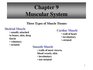
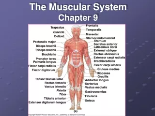
Skeletal Muscle Structure • Tendon – • Fascia – outermost covering; covers entire muscle & continuous w/tendon; separates muscle from adjacent muscles • Aponeuroses-
Skeletal Muscle Structure Coverings: • Epimysium – covers entire muscle (under fascia) • Perimysium – • Endomysium – covers each fiber (cell) • Sarcolemma –
Skeletal Muscle Structure Skeletal Muscle Structure – Cont. • Sarcoplasmic reticulum (SR) channels for transport • Myofibrils – threads that compose muscle fibers; contain protein filaments: 1. actin – 2. myosin –
Muscle Fiber(muscle cell) • Cisternae of SR – enlarged portions • Transverse tubules (T-tubules) – important in muscle contraction • Sarcoplasm –
Parts of a Sarcomere • Z lines – end points • M line – • I band – on either side of Z line; actin filaments only • H zone – on either side of M line; myosin filaments only • A band –
Troponin & Tropomyosin • 4 proteins are found in muscle cells: actin, myosin, troponin & tropomyosin • troponin – appear as globules; provide a binding site for Ca+² • tropomyosin–
Sliding Filament Theory (How Muscles Contract) • Muscle fiber stimulated by release of ACh from synaptic vesicles of neuron • Transverse tubules (T-tubules) carry impulse deep into muscle fibers • Ca²+ bind to troponin, tropomyosin moves, exposing binding sites on actin filaments
Cross Bridge Animation • cross bridge animation
Sliding Filament Theory (How Muscles Contract ) • Linkages form b/t actin & myosin • Actin filaments move inward, shortening the sarcomere • The enzyme cholinesterase (or AChesterase) decomposes ACh
Sliding Filament Theory • Relaxed muscle – binding sites on actin are covered by tropomyosin
Sliding Filament Theory • Ca²+ binds to troponin • Tropomyosin slides out of the way • Myosin binds to actin & pulls inward • Sarcomeres shorten & muscle contracts
Sliding Filament Animation • sliding filament animation
Neuromuscular Junction – junction b/t motor neuron & muscle • Motor neuron • Motor end plate – end of muscle fiber; many nuclei & mitochon- dria located here
Neuromuscular Junction • Neurotransmitters (ntm) chemicals that help carry impulses • Motor unit – • Synaptic vesicles – store neurotransmitter; most common – acetylcholine (ACh)
Neuromuscular Junction Animation • Neuromuscular Junction Animation
Energy for Muscle Contraction • ATP (adenosine triphosphate) • When ATP is converted to ADP(adenosine diphosphate) by losing the last phosphate, energy is released.
Energy for Muscle Contraction • Cells depend on cellular respiration of glucose to synthesize ATP • An additional source is creatine phosphate
Energy for Muscle Contraction • Creatine phosphate stores excess energy • Anaerobic respiration (in the absence of O2) provides few ATP’s, while aerobicresp. (in the presence of O2) provides many ATP’s
Creatine Phosphate High amts. of ATP - ATP is used to Low amts. of ATP – CP is used synthesize CP, which stores energy to resynthesize ATP. for later use.
Importance of Myoglobin • l.a. carried by blood to liver; liver can convert l.a. to glucose, but requires ATP (ATP being used for muscle contraction) • myoglobin –
Aerobic vs. Anaerobic Respiration Carried by blood to liver; liver can convert l.a. to glucose, but requires ATP (ATP being used for muscle contraction) Imp. b/c blood supply during muscle contr. may decrease As l.a. accumulates, O2 debt occurs
Oxygen Debt • Strenuous exercise leads to O2 deficiency & lactic acid buildup • ATP provides energy for muscle contraction • Amt. of O2 needed to convert accumulated l.a. to glucose & restore ATP levels = • L.A. accumulation leads to muscle fatigue b/c pH of muscle cell is lowered & muscle cannot contract
Muscle Cramp • Muscle cramp – • Rigor mortis – takes up to 72 hrs. to occur; sarcolemma becomes more permeable to Ca+² & ATP levels insufficient
Myogram Pattern or graph of a muscle contraction A single contraction is called a 3 parts: Latent (lag) phase – brief pd. of delay b/t when the stimulus is applied & actual contraction occurs Contraction Relaxation –
Patterns of Contraction • a) MuscleTwitch – • b) Staircase Effect many stimuli closely spaced w/complete relaxation in b/t; each contraction generate incr. force
Patterns of Contraction • c) Summation – when the 2nd stimulus occurs during the relaxation pd. of 1st contr.; the 2nd contr. generates more force • d) Tetany –
Muscle Facts • If a muscle is stimulated twice in quick succession, it may not respond the 2nd time – called refractory period • Threshold – • All-or-none – increasing the strength of the stimulation does NOT incr. the degree of contraction (a muscle contracts completely or not at all)
More Facts • Incr. stimulation from motor neurons causes a greater # of motor units to contract & vice versa • Called recruitment of motor units • Incr. the rate of stimulation also incr. the degree of contraction • Muscle tone –
Hypertrophy vs Atrophy • Hypertrophy- with intense exercise where muscle exerts more than 75% of its max tension there is an increase in actin and myosin fibers and increase muscle fiber diameter. • Inc diameter= • Atrophy-
Origin & Insertion • Origin – end of muscle that attaches to stationary bone • Insertion – • During contr., insertion is pulled toward origin
Muscle Functions in Groups Prime mover – responsible for most of the movement (ex.- biceps) Synergist – aids the prime mover Antagonist –
Fast vs Slow Twitch Fibers • Fast vs Slow Twitch Video