Download
1 / 77
780 likes | 1.31k Views
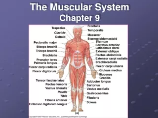
Chapter 9 Muscular System. Three Types of Muscle Tissues. Skeletal Muscle usually attached to bones under conscious control striated. Cardiac Muscle wall of heart not under conscious control striated. Smooth Muscle walls of most viscera, blood vessels, skin

E N D
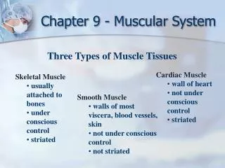
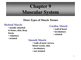
Chapter 9Muscular System Three Types of Muscle Tissues Skeletal Muscle • usually attached to bones • under conscious control • striated Cardiac Muscle • wall of heart • not under conscious control • striated Smooth Muscle • walls of most viscera, blood vessels, skin • not under conscious control • not striated
Muscle Tissues Skeletal Muscle Smooth Muscle Cardiac Muscle
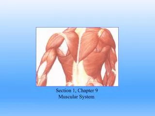
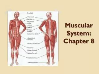
Structure of a Skeletal Muscle Skeletal Muscle • organ of the muscular system - skeletal muscle tissue - nervous tissue - blood - connective tissues • fascia • tendons • aponeuroses
Connective tissue coverings • Fascia (dense connective tissue) covers each skeletal muscle. - may project beyond end of muscle to form a cordlike tendon (muscle to bone) • Other connective tissues form broad fibrous sheets (aponeuroses) which may attach to coverings of adjacent muscles. • Epimysium - layer that closely surrounds skeletal muscle
Perimysium - layer that extends inward and separates muscle tissue into small sections • Fascicles - bundles of skeletal muscle fiber in the small sections • Endomysium - thin covering than surrounds each muscle fiber within a fascicle • Layers of connective tissue enclose and separate all parts of skeletal muscle allowing somewhat independent movement.
Fascia is a complex network that extends throughout the body • Deep fascia - surrounds each muscle • Subcutaneous fascia - lies just beneath skin • Subserous fascia - covers serous membranes
Skeletal muscle fibers • Each skeletal muscle fiber is a single muscle cell. • Fibers are cylindrical with rounded ends that are attached to connective tissues • Each cell has many nuclei • Muscle cell membrane - sarcolemma
Cytoplasm = sarcoplasm The sarcoplasm contains: • nuclei - control center • mitochondria - site of cellular respiration • sarcoplasmic reticulum - membraneous channels that activate muscle contraction when stimulated
cisternae - enlarged portion of sarcoplasmic reticulum • transverse tubules - membraneous channels that activate myofibrils (threadlike protein filaments) actin -thin filaments myosin - thick filaments
Actin and Myosin • arrangement of the filaments causes striations: • I bands(light) - thin actin • Z lines - I bands attach to it • A bands( dark) - thick myosin overlapping thin actin • myosin attached to Z lines by titin (protein) • H zone - central region of only thick filaments (myosin) • M line - thickening area on H zone • Sarcomere - extends from one Z line to the next Z line
Sarcomere • I bands • A bands • H zone • Z lines • M line
Neuromuscular Junction • Each muscle fiber is connected to an extension of a motor neuron • extends from the brain or spinal cord • Motor neurons stimulate muscle fibers to contract • Nerve fiber and muscle fiber meet at the neuromuscular junction • The motor end plate lies on one side of a neuromuscular junction
Synaptic cleft separates the nerve fiber and muscle fiber • In response to a nerve impulse, the end of a motor nerve fiber secretes a neurotransmitter which diffuses across the junction and stimulates the muscle fiber to contract.
Motor units • Made of one motor neuron and the muscle fibers associated with it • Number of muscle fibers varies • Fewer muscle fibers = finer movements Ex: eyes • More muscle fibers = coarse movements Ex: large muscle in the back
Motor Unit • single motor neuron • all muscle fibers controlled by motor neuron
Role of myosin and actin • Muscle fiber contraction results from a sliding movement of actin and myosin filaments. • About 2/3 of protein in skeletal muscle is myosin • Myosin consists of 2 twisted protein strands with globular cross bridges projecting outward
If Ca2+ present cross-bridges of myosin filaments form linkages with actin filaments. • Actin - myosin interactions provides the basis for contraction in all 3 • muscle types. • Actin is about 1/4 of the protein in skeletal muscle • Many actin molecules aggregate into double twisted strand
When a fiber is at rest, proteins troponin and tropomyosin molecules interfere with linkage formation. • Ca ions remove the inhibition, binding to troponin and altering the position of tropomyosin exposing the binding sites on actin.
Sliding filament theory • When stimulated to contract the head of a myosin cross bridge attaches to actin binding site, pulling the actin filament toward the center of the sarcomere, shortening the muscle • ATP converted to ADP releasing energy
Stimulus for contraction • Muscle fiber is usually stimulated by acetylcholine (ACh) • released from the end of a motor nerve fiber. • Ach is synthesized in cytoplasm of a motor neuron • ACh combines with receptors in the sarcolemma and stimulates it
A muscle impulse travels through the transverse tubules to the sarcoplasmic reticulum (SR) which has a high concentration of Ca2+ • cisternae membranes become more permeable to Ca2+ diffuse out of cisternae into the sarcoplasm causing linkages to form between actin and myosin which leads to muscle contraction
Contraction continues as long as ATP and ACh are present • Cessation of nerve impulse leads to muscle relaxation • ACh is decomposed by acetylcholinesterase • Stimulus to sarcolemma ceases, Ca2+ goes back into SR - Cross bridge linkages break and recock • Muscle relaxes
Energy sources for contraction • ATP supplies the energy for muscle fiber contraction • Muscles only have enough ATP to contract briefly • ATP must be regenerated • creatine phosphate stores energy that can be used to synthesize ATP as it is decomposed
The amount of ATP and creatine phosphate in a muscle can sustain contraction for only a few seconds • Active muscles depend upon cellular respiration of glucose for energy • Muscles store glucose in the form of glycogen
Anaerobic respiration yields few ATP molecules (2), whereas aerobic respiration provides many ATP's (34) • Hemoglobin in red blood cells carries oxygen from the lungs to body cells
Myoglobin(pigment) in muscle cells stores some oxygen temporarily reducing a the need for a continuous blood supply during contraction • Contracting muscle fibers may compress blood vessels
Oxygen debt • During rest or moderate exercise, oxygen is sufficient to support aerobic respiration • During strenuous exercise, an oxygen deficiency may develop, and lactic acid may accumulate as a result of anaerobic respiration • Lactic acid is carried by blood to the liver where it can be converted to glucose
The amount of oxygen needed to convert accumulated lactic acid to glucose and to restore supplies of ATP and creatine phosphate is called oxygen debt • Several hours may be needed to repay an oxygen debt after strenuous exercise
Muscle fatigue • A fatigued muscle loses its ability to contract • Usually due to the effects of accumulation of lactic acid • Lactic acid lowers the pH and prevents muscle fibers from responding to stimulation
Muscle cramps are due to a lack of ATP • ATP moves Ca2+ into SR and breaks the linkages between actin and myosin so the muscle fibers can relax • In a cramp part of the muscle may contract uncontrollably and other parts may be rigid
Physical training promotes new capillary growth and more mitochondria within muscles • Athletes usually produce less lactic acid than nonathletes because of their increased ability to supply oxygen and nutrients to muscles
Fast and slow muscles • The speed of contraction is related to a muscle's specific function • Slow-contracting(slow twitch) muscles contain oxygen storing myoglobin (red pigment) and many mitochondria • - can generate ATP fast enough to keep up with ATP breakdown and can contract for long periods Ex: long back muscles
Fast-contracting(fast twitch) muscles have less myoglobin, a poorer blood supply and fewer mitochondria - have reduced ability to carry on aerobic respiration and tend to fatigue relatively rapidly Ex: hand and eye muscles • Most skeletal muscles have both types
Heat production • By-product of cellular respiration • Active muscles release large amounts of heat • Blood transports heat throughout the body
Muscular Responses Threshold stimulus • Minimal strength of stimulation needed to cause a muscular contraction . All-or-none response • A muscle fiber does not partially contract • If a muscle fiber contracts at all, it will contract completely.
Recording a muscle contraction • A myogram is a recording of an electrically stimulated isolated muscle pulling a lever.
A twitch is a single contraction lasting a fraction of a second • The latent period is the time between stimulus and responding muscle contraction (0.01 second in a frog) • The refractory period immediately follows a contraction - a muscle cannot respond (very brief)