Download
1 / 17
180 likes | 208 Views
Learn about cartilage, its components (cells, matrix, perichondrium), three types (hyaline, elastic, fibrocartilage), growth methods, and limited repair ability.

E N D
Cartilage Dr. Ahmad Al-Taib Written by: Albara Marwa,, Abo Malik Group A leader
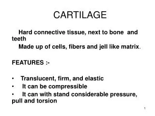
What is cartilage? • It is a special type of connective tissue with a firm extracellular matrix.
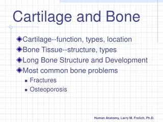
Location • Cartilage is found in: • Respiratory tract • External ear • Intervertebral disc • Articular surface of bones (in joints) • Skeleton of fetus
Components • Cartilage consists of three components: • Cells • Matrix (between the cells) • Perichondrium (outer layer)
Cartilage cells • Cartilage contains three types of cells: • Chondrogenic cells • Chondroblast • chondrocyte
1. Chondrogenic cells • Originate from mesenchymal cells • Spindle shaped cells with oval nuclei • Found in the perichondrium • Differentiate into chondroblasts (Differentiation of cells does not include cell division but only a change in structure)
2.Chondroblasts • Basophilic flat cells with flat nuclei • Protein-secreting cells • Secrete cartilage matrix • Found in the perichondrium • Originate from chondrogenic cells
3. Chondrocyte • Each chondroblast secretes the matrix & becomes a chondrocyte with a large rounded nucleus. • The chondrocyte is the mature cartilage cell which also secretes matrix. • Each chondrocyte is situated in a small space called a lacuna surrounded by the matrix. • The chondrocyte can divide in the lacuna • There is no connection between the lacunae.
Cartilage Matrix • The matrix is an avascular extracellular material secreted by chondroblast & chondrocyte. • Cartilage matrix consists of: • Fibers: collagen type I or type II or elastic fibers. • Ground substance: glycoproteins & chondroitin sulphate.
Perichondrium • A dense vascular connective tissue on the surface of cartilage. • It consists of: • Outer fibrous layer of dens CT (Type I collagen). • Inner cellular layer of chondrogenic cells & chondroblast. • Its function is growth of cartilage
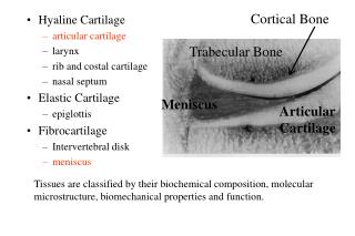
Types of cartilage • There are three types according to the fibers in the matrix: • Hyaline cartilage • Elastic cartilage • Fibrocartilage
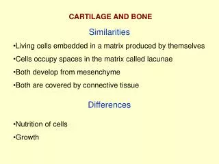
Similarities between types of cartilage • All types have chondrocyte in lacunae. • All types have avascular matrix. • All types have perichondrium EXCEPT: • Fibrocartilage • Articular cartilage
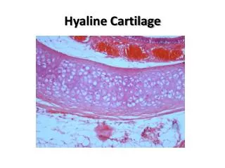
Hyaline cartilage • Found in: • Respiratory tract • Articular cartilage • Chondrocytes divide and form small groups called isogenous group. • Collagen type II in a basophilic matrix, but the collagen is not visible with the L.M. • The matrix is more basophilic near the chondrocyte. • Articular cartilage lacks perichondrium and it receives nutrient from the synovial fluid
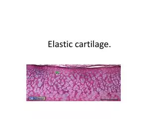
Elastic cartilage • More chondrocytes and less matrix than hyaline cartilage. • Many elastic fibers in the matrix and in the perichondrium. • Collagen type II is also present • Examples: External ear & epiglottis. • Elastin stain stains elastin black
Fibrocartilage • Parallel bundles of acidophilic type I collagen fibers in the matrix. • (Note: Collagen type I is stronger than type II). • Chondrocytes form parallel rows. • Chondrocytes arise from fibroblasts. • More fibers and less chondrocytes than that in hyaline and elastic cartilage. • No perichondrium • Example: Intervertebral disc
Cartilage growth • Cartilage arises from mesenchyme during chondrogenesis • Cartilage grows by two methods: • Appositional growth : on surface from perichondrium. • Interstitial growth: deep in matrix.
Repair of cartilage • Cartilage has a limited ability for repair. • Injured cartilage is replaced by Connective tissue.