Download
1 / 34
340 likes | 438 Views
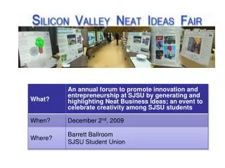
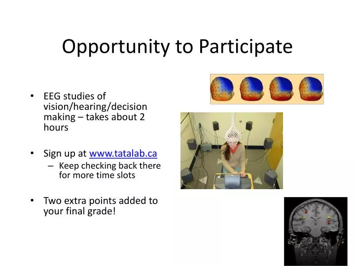
Opportunity to Participate. EEG studies of vision/hearing/decision making – takes about 2 hours Sign up at www.tatalab.ca Keep checking back there for more time slots Two extra points added to your final grade!. Functional Imaging.

E N D
Opportunity to Participate • EEG studies of vision/hearing/decision making – takes about 2 hours • Sign up at www.tatalab.ca • Keep checking back there for more time slots • Two extra points added to your final grade!
Functional Imaging • Oxygenated hemoglobin is diamagnetic - it has no magnetic effects on surrounding molecules • Deoxygenated hemoglobin is paramagnetic - it has strong magnetic effects on surrounding molecules! Hemoglobin
Functional Imaging • blood flow overshoots baseline after a brain region is activated • More oxygenated blood in that region increases MR signal from that region
Principles of MRI • Some terms: • Nuclear Magnetic Resonance (NMR) • quantum property of protons • energy absorbed when precession frequency matches radio frequency • Magnetic Resonance Imaging (MRI) • uses spatial differences in resonance frequencies to form an image • basis of anatomical MRI • functional Magnetic Resonance Imaging (fMRI) • exploits magnetic properties of hemaglobin to create images changes in cortical blood flow
Principles of MRI • Some terms: • Nuclear Magnetic Resonance (NMR) • quantum property of protons • energy absorbed when precession frequency matches radio frequency • Magnetic Resonance Imaging (MRI) • uses spatial differences in resonance frequencies to form an image • basis of anatomical MRI • functional Magnetic Resonance Imaging (fMRI) • exploits magnetic properties of hemaglobin to create images changes in cortical blood flow
Principles of MRI • Some terms: • Nuclear Magnetic Resonance (NMR) • quantum property of protons • energy absorbed when precession frequency matches radio frequency • Magnetic Resonance Imaging (MRI) • uses spatial differences in resonance frequencies to form an image • basis of anatomical MRI • functional Magnetic Resonance Imaging (fMRI) • exploits magnetic properties of hemaglobin to create images changes in cortical blood flow
Principles of MRI • Some terms: • Nuclear Magnetic Resonance (NMR) • quantum property of protons • energy absorbed when precession frequency matches radio frequency • Magnetic Resonance Imaging (MRI) • uses spatial differences in resonance frequencies to form an image • basis of anatomical MRI • functional Magnetic Resonance Imaging (fMRI) • exploits magnetic properties of hemaglobin to create images changes in cortical blood flow
Principles of NMR • Protons are like little magnets • they orient in magnetic fields like compass needles • what way do they normally point?
Principles of NMR • Protons are like little magnets • they orient in magnetic fields like compass needles • what way do they normally point? • normally aligned with Earth’s magnetic field
Principles of NMR • Protons are like little magnets • they orient in magnetic fields like compass needles • what way do they normally point? • normally aligned with Earth’s magnetic field • NMR uses a big magnet to align all the protons in a sample (e.g. brain tissue)
Principles of NMR • Protons are like little magnets • Radio Frequency pulse will knock protons at an angle relative to the magnetic field
Principles of NMR • Protons are like little magnets • Radio Frequency pulse will knock protons at an angle relative to the magnetic field • once out of alignment, the protons begin to precess
Principles of NMR • Protons are like little magnets • Radio Frequency pulse will knock protons at an angle relative to the magnetic field • once out of alignment, the protons begin to precess • protons gradually realign with field (relaxation)
Principles of NMR • Protons are like little magnets • Radio Frequency pulse will knock protons at an angle relative to the magnetic field • once out of alignment, the protons begin to precess • protons gradually realign with field (relaxation) • protons “echo” back the radio frequency that originally tipped them over • That radio “echo” forms the basis of the MRI image
Principles of NMR • Protons are like little magnets • The following simple equation explains MRI image formation
Functional Imaging • Recall that precessing protons give off a radio “echo” as they realign with the magnetic field • We pick up the combined echo from many protons that are in phase
Functional Imaging • recall that the precession frequency depends on the field strength • anything that changes the field at one proton will cause it to de-phase
Functional Imaging • recall that the precession frequency depends on the field strength • anything that changes the field at one proton will cause it to de-phase • The de-phased region will give off less echo
Functional Imaging • It is important to recognize that fMRI “sees” changes in the ratio of oxygenated to deoxygenated blood - nothing more • BOLD: Blood Oxygenation Level Dependant contrast
Functional Imaging • How do we create those pretty pictures? • We ask the question “When the subject engages in this cognitive task, where does blood oxygenation change significantly?” “where does it change randomly?”
Experimental Design in fMRI • Experimental Design is crucial in using fMRI • Simplest design is called “Blocked” • alternates between active and “rest” conditions Active Rest Active Rest 60 sec 60 sec 60 sec 60 sec
Experimental Design in fMRI • Experimental Design is crucial in using fMRI • Simplest design is called “Blocked” • alternates between active and “rest” conditions Active Rest Active Rest 60 sec 60 sec 60 sec 60 sec
Experimental Design in fMRI • A voxel in tissue insensitive to the task demands shows random signal change Signal Active Rest Active Rest 60 sec 60 sec 60 sec 60 sec
Experimental Design in fMRI • A voxel in tissue that responds to the task shows signal change that matches the timecourse of the stimulus Signal Active Rest Active Rest 60 sec 60 sec 60 sec 60 sec
Experimental Design in fMRI • A real example of fMRI block design done well: • alternate moving, blank and stationary visual input Moving Blank Stationary Blank 40 sec 40 sec 40 sec 40 sec
Experimental Design in fMRI • Voxels in Primary cortex tracked all stimuli
Experimental Design in fMRI • Voxels in area MT tracked only the onset of motion
Experimental Design in fMRI • Voxels in area MT tracked only the onset of motion • How did they know to look in area MT?
PET: another way to measure blood Oxygenation • Positron Emission Tomography (PET) • Injects a radioisotope of oxygen • PET scanner detects the concentration of this isotope as it decays
Advantages of fMRI • Advantages of MRI: • Most hospitals have MRI scanners that can be used for fMRI (PET is rare) • Better spatial resolution in fMRI than PET • Structural MRI is usually needed anyway • No radioactivity in MRI • Better temporal resolution in MRI
Advantages of PET • Advantages of PET: • Quiet • A number of different molecules can be labeled and imaged in the body
Limitations of fMRI • All techniques have constraints and limitations • A good scientist is careful to interpret data within those constraints
Limitations of fMRI • Limitations of MRI and PET: • BOLD signal change does not necessarily mean a region was specifically engaged in a cognitive operation • Poor temporal resolution - depends on slow changes in blood flow • expensive