Download
1 / 49
490 likes | 584 Views
Understand neoplastic eye diseases, from eyelid tumors to malignant melanoma of the uvea. Learn about diagnosis, treatment, and monitoring methods. Stay informed to protect your vision.

E N D
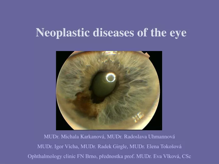
Neoplastic diseases of the eye MUDr. Michala Karkanová, MUDr. Radoslava Uhmannová MUDr. Igor Vícha, MUDr. Radek Girgle, MUDr. Elena Tokošová Ophthalmology clinic FN Brno, přednostka prof. MUDr. Eva Vlková, CSc
Tumor tissue change, which is a result of the locallynoncontrolablegrowth of autonomous nature. The biological nature of the tumor: benign malignant Separation of eye tumors according to anatomiclocalization: eyelid tumorstumors of the eye orbital tumors
Location:anywhere on the capmainly a cosmetic problemfault status and function lids with symptoms of dry eye syndrome (burning, cutting, more frequent sec. infections, xerosis of the conjunctiva, exposure keratopathy a reduction or even loss of the eye ZO)Treatment:(Depending on size, location and nature of the changes) Early excision with a sufficiently large safety rim histological verification Eyelidstumors
Location:anywhere on the lid, withoutagelimitationmostly a cosmeticproblemRetentioncystssebaceousglands (milium, atheroma)Papilloma - cutaneoushornsVerruca, verrucasenileHemangiomaXantelasmaNevusTreatment:Observation (nevi)Surgery - cautery, simpleexcision, laser therapy (CO2 laser), cryoHistologicalexamination !!! Benigneyelidstumors
Benigneyelidstumors Eyelids papiloma Retention cyst
Location: predilectivelylower lid, 6.-7. decadeoflife basal cell carcinoma (invasiononlylocal) squamous cell carcinoma(metastasizes) malignantmelanoma MeibomglandscarcinomaTreatment: surgicalexcision - simple - withplasticfinish radiotherapy surgeryfollowed by radiotherapy localapplication ILOncologicdispensary! Malignanteyelidtumors
Basal cell carcinoma Malignanteyelidtumors
Location: • predilectively range of eye slits, all ages, a shift to a higher ageTreatment:dispensary congenital change without progression -photographs (cosmetic point of view)surgical- block excision, lamellar keratectomy,in malignancies completed withcryotherapy-radical excision (up orbit exenteration)additional local radiotherapylocal applicationofantimetabolitesHistological examination!Oncological dispensary in melanoma and cancer! • Lokalizace: • predilekčně rozsah oční šterbiny, všechny věkové kategorie • s posunem do vyššího věku • Léčba: • dispenzarizace vrozených změn bez progrese - • fotodokumentace ( kosmetické hledisko ) • chirugická - excize bloková, lamelárníkeratektomie, • u malignit doplněno kryalizací • - excise radikální (až exenterace očnice) • doplňková lokální radioterapie • lokápní aplikace antimetabolitů • Histologické vyšetření ! • Onkologická dispenzarizace v případě melanomu a karcinomu! Tumorsoftheconjunctiva and cornea
Congenital: Choristoma - dermoid, lipodermoid HemangiomaEpithelial: Hyperplasia Epithelioma (carcinoma in situ, Bowen'sdisease) Melanotic: Melanosis - congenital - acquired (withorwithoutatypiaatypical) Nevus, Melanocytoma (kong. based) Benigntumorsoftheconjunctiva and cornea
conjunctival papiloma conjunctival lipodermoid Benigntumorsoftheconjunctiva and cornea conjunctival lymfangioma
conjunctival nevus carcinoma in situ Benigntumorsoftheconjunctiva and cornea conjunctival melanosis
Malignant melanoma of the conjunctiva • Carcinoma of the conjunctiva ( rare disease)) • Lymfoma of the conjunctiva (Non – Hodgkin type) Malignanttumorsoftheconjuntiva and cornea conjunctival lymfoma conjunctival malignant melanoma
Primary: the origin of the uvea (iris, ciliary body, choroid) originate in the retina (exceptionally on adults)Secondary: infiltrative growth of surrounding tissueMetastatic: following generalization of the malignancy most common in the choroid (often the first symptom of malignancy) Metastases - women breast carcinoma 85%, bronchi8% - male lung carcinoma 38%, GIT 20% Intraoculartumors
Iris 8% Ciliary body 12% Chorioid 80% the most common primary intraocular tumor of adults incidence between 50-70 years featured mortality 30 -70%most often unilateral Malignantmelanomaofthe uvea( MMU )
MMU Diagnostics Examination on the slit lamp Ophthalmoscopy • direct • indirect • biomicroskopye • gonioscopy Sonography • B scan • standard. echography • UBM
MMU Diagnostics FAG ( fluorescein angiography ) ICG ( indocyaninangiography ) CT, NMR
Examinations performed in determining the MMU diagnosis • Completlaboratoryexaminationsincludingoncomarkers and melanogensin urine • Lungs radiology • Echographyofparenchymatousorgansofthe abdomen • Sceletonscintigraphy • Brain CT , NMR in suspect. metastasis • Completinnerexaminatin • Onkologicalexaminatin • ( PET )
Criteria for selecting therapeutic approach • individual • vision, intraoculartension, status oftheaffectedeye • sizeofthe tumor, signsofitsactivities • localization, shape • othereyecondition, pacientsgeneralstate • ageofthepatientatthetimeofdetection
Iris malignantmelanoma • most common occurrence in the lower half of the iris • variouspigment • distortion of the pupil • ectopiaofpigmentedsheet • partial cataract
Diferencialdiagnosisofthe iris tumors • nevus • cyst • leiomyoma • leaf pigment hyperplasia iris like the tiger nevus of the iris
Treatment of benign and malignant lesions of the iris • monitoring borderlinefindings (photographs) • excision - in suspectedlesionsnotoverlaping 4 hours • enucleationofthe globe - susp. malignantlesionsover 1/2 ofthe iris, blind bulb, noncorrectedsecondaryglaucoma
Ciliary body malignantmelanoma • long asymptomatic • extensionepiscleralvessels • pressureon thelens (astigmatism, partialcataract, subluxation) • secondaryretinaldetachment • iris rooterosion • secondaryglaucomaafterinitialhypotension • epibulbarmeat in place ofextrabulbarextension
Diferencialdiagnosisofciliary body tumors • tumorsfromthe pigment and nonpigmentepithelium • cysts • clinicalindistinguishable cyst of ciliary body
Therapyofciliary body melanomas • cyclectomy • iridocyclectomy • radiotherapy - brachytherapy Lexell gama knife • enucleation
Diferencialdiagnosisofchoroidallesions • exudativeformof ARMD • chorioidalgranulomatousscars • subretinalhaemorrhage • big prominent nevi • hyperplasiaof RPE • ablationofthechoroid • metastases • cavernoushemangioma • rearscleritis • melanocytoma • retinoblastoma
HistologicalclassificationaccordingCallender • spindle type A • spindletype B • epithelioid • mixed • fascicular
Spindle type A: mortality 5% in 5 years Spindle type B: 14% in 5 years Epithelioid type: 69% in 5 years Necrotic type: untill 50% in 5 years Prognosticfactors MM cell type size localization Bruchmembranestate extrabulbarextension Prognosisquadvitamaccordinghistological type ofthe tumor:
Metastases At the time of finding the MMU has about 11% of metastases simultaneously. Most commonlocalization and % behalf: • liver 60-70 • subcutaneus 24 • lungs 7 • spine 7 • CNS 2
Nonactivelesions inaccuratelybounded occurrenceofdrusen on thesurface Activelesions documentedgrowth ( measured by ultrasound) boundedelevation breakingBruchsmembrane productionof SRF occurrenceoflipofuscin on surfaceofthetunmor Signsof tumor activity
Sizeofthe tumor – classification by Shields • melanomas to 3mm • melanomas to 5mm • melanomas to 10mm • melanomasabove 10mm
Therapyofchoroidal MM • Photocoagulation • TTT • Photodynamictherapy • Radiotherapy • Brachytherapy • Lexell gama knife • Parcialresectionofthe tumor • Enucleationofthebulb • Exenterationofthe orbit
Brachytherapy Indication • Height to 10 mm • Bases to 15 mm radioactive source 106Ru
Enucleation of the bulb • heightabove 8-10 mm • basesabove 15 mm • smallrangeextrabulbarextension • blind and painfullbulbswithsecondaryglaucoma
Exenterationofthe orbit Indications: • retrobulbarextensionofthe tumor • significantperibulbarextensionofthe tumor
Dispenzary In a subsequent patient care is extremely important collaboration between an ophthalmologist, internal physician and oncologist who will decide on possible further therapy (cytostatics, interferon ...).
Závěr • V přednášce byly použity materiály a obrazová dokumentace z následujících knih a sdělení: • Nádory oka a očních adnex u dospělých, MUDr. Radoslava Uhmannová, • III. celostátní sjezd oftalmologické sekce České asociace sester,10/ 2006, Brno • Nádory oka, Prof. MUDr. Drahomíra Baráková, CSc. a kol., Praha 2002 • Maligní melanom uvey ( současná diagnostika a léčba ), MUDr. R. Girgle, • MUDr. Radoslava Uhmannová, MUDr. Igor Vícha • Enukleace bulbu, Eviscerace bulbu, Exenterace očnice, MUDr. Igor Vícha, • MUDr. Radoslava Uhmannová, MUDr. Michala Karkanová • Závěrem děkuji všem zmíněným autorům za poskytnutí jejich materiálů a všem lékařům Oční kliniky FN Brno za poskytnutí obrazové dokumentace.