Download
1 / 2
20 likes | 121 Views
Supplemental Figure 2. a. ADP titration (Clark-type electrode). ~10.00. ~10.27. ~7.01. ~5.11. ~5.00. ~4.03. ADP titration (Seahorse XF24). b. ~3.89. ~3.10. ~2.62. ~2.03. ~1.63. c. Mitochondrial integrity test (Clark-type electrode). d.

E N D
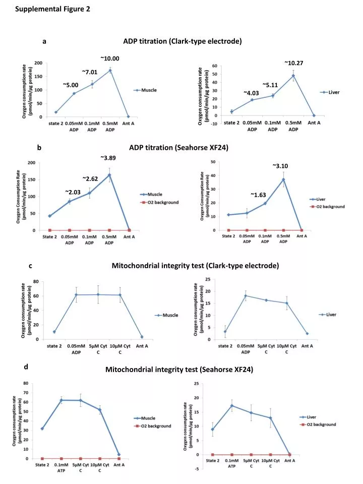
Supplemental Figure 2 a ADP titration (Clark-type electrode) ~10.00 ~10.27 ~7.01 ~5.11 ~5.00 ~4.03 ADP titration (Seahorse XF24) b ~3.89 ~3.10 ~2.62 ~2.03 ~1.63 c Mitochondrial integrity test (Clark-type electrode) d Mitochondrial integrity test (Seahorse XF24)
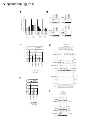
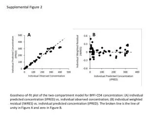
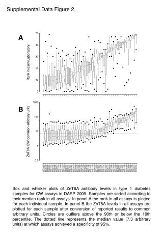
Figure S2: Validation of Seahorse XF24 method for measuring respiration in isolated mitochondria. A) Assessment of respiratory control ratio (RCR) by Clark-type electrode in muscle (left panel) liver (right panel) mitochondria. Following measurement of state 2, ADP was sequentially injected to a final concentration of 0.5 mMto test state 3 respiration rates. Antimycin A (Ant A; 10 μM) was then injected to inhibit respiration. B) Assessment of RCR by Seahorse XF24 in muscle (left panel) liver (right panel) mitochondria. Following measurement of state 2, ADP was sequentially injected to a final concentration of 0.5 mM.Antimycin A (Ant A; 4 μM) was then injected. RCR was estimated as the ratio of state 3/state 2. n=2, mean±SEM. C) Assessment of mitochondrial integrity by Clark-type electrode in muscle (left panel) liver (right panel) mitochondria. Following measurement of state 2, state 3 respiration was stimulated using ADP (final concentration of 0.05 mM) followed by treatment with cytochrome C (final concentration of 10 μM). Antimycin A (Ant A; 10 μM) was then injected to deactivate respiration. D) Assessment of mitochondrial integrity by Seahorse XF24 in muscle (left panel) andliver (right panel) mitochondria. Following measurement of state 2, state 3 respiration was stimulated using ADP (final concentration of 0.1 mM) followed by treatment with sequential treatment with cytochrome C (final concentration of 10 μM). Antimycin A (Ant A; 4 μM) was then injected. n=2, mean±SEM.