Download
1 / 26
300 likes | 411 Views
Learn about types, causes, diagnosis, and treatment of impetigo, abscesses, cellulitis, and erysipela. Understand bacteria involved, diagnostic methods, and antimicrobial susceptibility.

E N D
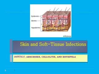
Skin and Soft-Tissue Infections IMPETIGO, ABSCESSES, CELLULITIS, AND ERYSIPELA
Objectives • Describe the anatomical structure of skin and soft tissues. • Differentiate the various types of skin and soft tissue infections and there clinical presentation. • Name bacteria commonly involved in skin and soft tissue infections • Describe the pathogenesis of various types of skin and soft tissue infections • Recognize specimens that are acceptable and unacceptable for different types of skin and soft tissue infections • Describe the microscopic and colony morphology and the results of differentiating bacteria isolates in addition to other non-microbiological investigation • Discuss antimicrobial susceptibility testing of anaerobes including methods and antimicrobial agents to be tested. • Describe the major approaches to treat of skin and soft tissue infections • either medical or surgical.
Introduction • Common • Can be mild to moderate or sever muscle or bone and lungs or heart valves . • Staphylococcus aureusis the most cause • Emerging antibiotic resistance among • Staphylococcus aureus (methicillin resistance) • Streptococcus pyogenes (erythromycin resistance)
key to developing an adequate differential diagnosis requires • History • patient’s immune status, the geographical locale, travel history, recent trauma or surgery, previous antimicrobial therapy, lifestyle, and animal exposure or bites • Physical examination • severity of infection • Investigation • CBCs, Chemistry • Swab, biopsy or aspiration • Radiographic procedures • Level of infection and the presence of gas or abscess. • Surgical exploration or debridement • Diagnostic and therapeutic
IMPETIGO-(Pyoderma) • A common skin infection • Children 2–5Yr in tropical or subtropical regions • Nearly always caused by β-hemolytic streptococci and/or S.aureusand / or Group A streptoccus • Nonbullous (Streptococcus) or Bullous (S. aureus) (Consists of discrete purulent lesions) • Exposed areas of the body( face and extremities) • Skin colonization- Inoculation by abrasions, minor trauma, or insect bites • Systemic symptoms are usually absent. • Poststreptococcalglomerulonephritis. • Cefazolin, Cloxacillin , or erythromycin • Mupirocin
ABSCESSES, CELLULITIS, AND ERYSIPELA • Cutaneous abscesses. • Collections of pus within the dermis and deeper skin tissues. • Painful, tender, and fluctuant • Typically caused by S. aureus • Do Gram stain, culture • Multiple lesions, cutaneous gangrene, severely impaired host defenses, extensive surrounding cellulitis or high fever. • and systemic antibiotics • Incision and evacuation of the pus
Furuncles and carbuncles. • Furuncles (or “boils”) are infections of the hair follicle (folliculitis ), usually caused by S. aureus, in which suppuration extends through the dermis into the subcutaneous tissue • Carbuncle- extension to involve several adjacent follicles with coalescent inflammatory mass - back of the neck especially in diabetics • Larger furuncles and all carbuncles require incision and drainage. • Systemic antibiotics are usually unnecessary
Outbreaks of furunculosis caused by MSSA, and MRSA, • Families-prisons-sports teams • Inadequate personal hygiene • Repeated attacks of furunculosis • Presence of S. aureus in the anterior nare- 20-40% • Mupirocin ointment- eradicate staphylococcal carriage nasal colonization
Erysipelas and Cellulitis. • Diffuse spreading skin infections • Most of the infections arise from streptococci, often group A, but also from other groups, such as B, C, or G. Erysipelas • Affects the upper dermis (raised-clear line of demarcation) • Red, tender, painful plaque • Infants, young children- • B-hemolytic streptococci ( group A or S. pyogenes.) • Penicillin-IV or oral.
Cellulitis • Acute spreading infection involves the deeper dermis and subcutaneous tissues. • β-hemolytic streptococci, Group A streptococci, and group B streptococci-diabetics • S. aureus : commonly causes cellulitis- penetrating trauma. • Haemophilusinfluenzaeperiorbitalcellulitisin children • Risk factors ; Obesity, venous insufficiency, lymphatic obstruction (operations), preexisting skin infections- ulceration, or eczema, • CA-MRSA • Carry Panton-Valentine leukocidin gene • More sensitive to antibiotics • Can lead to sever skin and soft tissue infection or septic shock
Diagnosis and Treatment • Clinical diagnosis Symptoms and Signs • High WBCs, blood culture rarely needed (Celullulitis) • Aspiration and biopsy , diabetes mellitus, malignancy, animal bites, neutropenia (Pseudomonas aeruginosa ) immunodeficiency, obesity and renal failure • progression to severe infection(increased in size with systemic manifestation. (fever, leukocytosis) • Treatment: cover streptococcus and staphylococcus • Penicillin, cloxacillin, cefazolin(cephalexin),clindamycin • Vancomycin or linazolid in case of MRSA • Clindamycin, TMP-SMZ for CaMRSA.
Necrotizing fasciitis flesh-eating disease
Introduction • rare deep skin and subcutaneous tissues infection • It can be monomicrobial or (polymicrobial) infection • Most common in the arms, legs, and abdominal wall and is fatal in 30%-40% of cases. • Fournier's gangrene (testicular), Necrotizing cellulitis • Group A streptococcus (Streptococcus pyogenes) • Staphylococcus aureus or CA-MRSA • Clostridium perfringens (gas in tissues) • Bacteroides fragilis • Vibrio vulnificus (liver function) • Gram-negative bacteria (synergy). • E. coli, Klebsiella, Pseudomonas • Fungi
Risk factors • Immune-suppression • Chronic diseases: ( diabetes, liver and kidney diseases, malignancy • Trauma:(laceration, cut, abrasion, contusion, burn, bite, subcutaneous injection, operative incision) • Recent viral infection rash (chickenpox) • Steroids • Alcoholism • Malnutrition • Idiopathic
Pathophysiology • destruction of skin and muscle by releasing toxins • Streptococcal pyogenicexotoxins • Superantigen • Non-specific activation of T-cells • Overproduction of cytokines • Severe systemic illness (Toxic shock syndrome)
Signs and symptoms • Rapid progression of sever pain with fever , chills (typical) • Swelling , redness, hotness, blister, gas formation, gangrene and necrosis • Blisters with subsequent necrosis , necrotic eschars • Diarrhea and vomiting (very ill) • Shock organ failure • Mortality as high as 73 % if untreated
A delay in diagnosis is associated with a grave prognosis and increased mortality • Clinical-high index of suspicion • Blood tests • CBC-WBC , differential,ESR • BUN (blood urea nitrogen) • Surgery debridement- amputation • Radiographic studies • X-rays : subcutaneous gases • Doppler CT or MRI • Microbiology • Culture &Gram's stain • ( blood, tissue, pus aspirate) • Susceptibility tests Diagnosis
Treatment • If clinically suspected patient needs to be hospitalized OR require admission to ICU • Start intravenous antibiotics immediately • Antibiotic selection based on bacteria suspected • broad spectrum antibiotic combinations against • methicillin-resistant Staphylococcus aureus(MRSA) • anaerobic bacteria • Gram-negative and gram-positive bacilli • Surgeon consultation • Extensive Debridement of necrotic tissue and collection of tissue samples • Can reduce morbidity and mortality
Treatment • Antibiotics combinations • Penicillin-clindamycin-gentamicin • Ampicillin/sulbactam • Cefazolin plus metronidazol • Piperacillin/tazobactam • Clostridium perfringens - penicillin G • Hyperbaric oxygen therapy (HBO) treatment
Pyomyositis • Acute bacterial infection of skeletal muscle, usually caused by Staph. aureus • No predisposing penetrating wound, vascular insufficiency, or contiguous infection • Most cases occur in the tropics • 60% of cases outside of tropics have predisposing RF: DM, EtOH liver disease, steroid rx, HIV, hematologic malignancy
Pyomyositis • Hx of blunt trauma or vigorous exercise (50%), then period of swelling without pain. 10-21 days later, pain, tenderness, swelling and fever, Pus can be aspirated from muscle. 3rd stage: sepsis, later metastatic abscesses if untreated • Dx: X-ray, US, MRI or CT • Rx: surgical drainage +abx
TAKE HOME POINTS • Most commonly caused by Staphylococcus aureus and Streptococcus pyogenes • Risk factors for developing SSTIs include breakdown of the epidermis, surgical procedures ,crowding, co-morbidities, venous stasis, lymph edema
TAKE HOME POINTS • Most SSTIs can be managed on an outpatient basis • patients with evidence of rapidly progressive infection, high fevers, or other signs of systemic inflammatory response should be monitored in the hospital setting. • Superficial SSTIs typically do not require systemic antibiotic treatment and can be managed with topical antibiotic agents or incision and drainage. • • Systemic antibiotic agents that provide coverage for both Staphylococcus aureus and Streptococcus pyogenesare most commonly used as empiric therapy for both uncomplicated and complicated deeper infections.