Download
1 / 72
920 likes | 3.06k Views
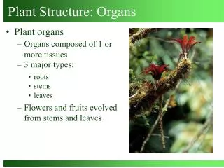
Plant Organs: Stems. Chapter 7. LEARNING OBJECTIVE 1. Describe three functions of stems. Stem Functions . Support leaves and reproductive structures Conduct water, dissolved minerals, carbohydrates Produce new living tissues at apical meristems

E N D
Plant Organs: Stems Chapter 7
LEARNING OBJECTIVE 1 • Describe three functions of stems
Stem Functions • Support • leaves and reproductive structures • Conduct • water, dissolved minerals, carbohydrates • Produce new living tissues • at apical meristems • at lateral meristems (secondary growth)
LEARNING OBJECTIVE 2 • Relate the functions of each tissue in an herbaceous stem
Tissues in Herbaceous Stems 1 • Epidermis • protective outer layer • covered by water-conserving cuticle • Vascular tissues • Xylemconducts water and dissolved minerals • phloemconducts dissolved carbohydrates (sucrose)
Tissues in Herbaceous Stems 2 • Storage tissues • Cortex and pith • ground tissue
LEARNING OBJECTIVE 3 • Contrast the structures of an herbaceous eudicot stem and a monocot stem
Herbaceous Eudicot Stems • Havevascular bundlesarranged in a circle (in cross section) • Have a distinct cortex and pith
Monocot Stems • Have scattered vascular bundles • Have ground tissue instead of distinct cortex and pith
Epidermis Ground tissue Vascular bundles Ground tissue Phloem Xylem Air space Bundle sheath Corn seedling (a) Cross section of a corn (Zea mays) stem, showing the scattered vascular bundles. (b) Close-up of a vascular bundle. The air space is where the first xylem elements formed. The entire bundle is enclosed in a bundle sheath of sclerenchyma for additional support. Fig. 7-4, p. 134
LEARNING OBJECTIVE 4 • Distinguish between the structures of stems and roots
Differences Between Stems and Roots 1 • Unlike roots, stems have nodesand internodes, leaves and buds • Unlike stems, roots have root caps and root hairs
KEY TERMS • NODE • Area on a stem where one or more leaves is attached; stems have nodes, but roots do not • INTERNODE • Stem area between two successive nodes
KEY TERMS • BUD • An undeveloped shoot that contains an embryonic meristem • May be terminal (at tip of stem) or axillary (on side of stem)
Terminal bud Bud scale One year’s growth Terminal bud scale scars Node Internode Axillary bud Leaf scar Node Lenticels Terminal bud scale scars Bundle scars Fig. 7-2, p. 131
Differences Between Stems and Roots 2 • Internally • herbaceous roots possess an endodermis and pericycle • stems lack a pericycle and rarely have an endodermis
LEARNING OBJECTIVE 5 • Outline the transition from primary growth to secondary growth in a woody stem • List the two lateral meristems, and describe the tissues that arise from each
Vascular bundles Pith Cortex Epidermis (a) Cross section of a sunflower (Helianthus annuus) stem, showing the organization of tissues. The vascular bundles are arranged in a circle. Sunflower Epidermis Cortex Phloem fiber cap Pith ray Phloem Vascular bundle Vascular cambium Xylem Vessel element Pith (b) Close-up of a vascular bundle. The xylem is toward the stem’s interior, and the phloem toward the outside. Each vascular bundle is “capped” by a batch of fibers for additional support. Fig. 7-3, p. 132
KEY TERMS • VASCULAR CAMBIUM • A lateral meristem that produces secondary xylem (wood) to the inside and secondary phloem (inner bark) to the outside
Secondary Growth • Occurs in woody eudicots and conifers • Produced by vascular cambium • between primary xylem and primary phloem
Vascular Cambium 1 • Is not initially a solid cylinder of cells • becomes continuous when production of secondary tissues begins
Vascular Cambium 2 • Certain parenchyma cells between vascular bundles • retain ability to divide • connect to vascular cambium cells in each vascular bundle • form a complete ring of vascular cambium
4X 2P 1P 3X 2X 1X 3X 2X 2P 1X 1P 1P 1X 2P 2X Time Secondary xylem Secondary phloem 1P 2X 1X Second division of vascular cambium forms a phloem cell. 1P 1X Division of vascular cambium forms two cells, one xylem cell and one vascular cambium cell. 1X Vascular cambium cell when secondary growth begins. Vascular cambium cell Fig. 7-6, p. 136
KEY TERMS • CORK CAMBIUM • A lateral meristem that produces cork parenchyma to the inside and cork cells to the outside • Cork cambium and the tissues it produces make up the outer bark of a woody plant
Cork Cambium • Arises near the stem’s surface • Is either a continuous cylinder of dividing cells or a series of overlapping arcs of meristematic cells that form from parenchyma cells in successively deeper layers of the cortex and, eventually, secondary phloem
Primary xylem Epidermis Cortex Primary phloem Vascular cambium Pith (a) At the onset of secondary growth, vascular cambium arises in the parenchyma between the vascular bundles (that is, in the pith rays), forming a cylinder of meristematic tissue (blue circle in cross section). Fig. 7-5a, p. 135
Remnant of cortex Remnant of epidermis Remnant of primary phloem Secondary phloem (inner bark) Secondary xylem (wood) Periderm (outer bark) Remnant of primary xylem Remnant of pith Vascular cambium (b) Vascular cambium begins to divide, forming secondary xylem on the inside and secondary phloem on the outside. Fig. 7-5b, p. 135
Periderm (outer bark; remnants of primary phloem, cortex, and epidermis are gradually crushed or torn apart and slough off) Secondary xylem (wood) Secondary phloem (inner bark) Remnant of primary xylem Remnant of pith Vascular cambium (c) A young woody stem. Vascular cambium produces more secondary xylem than secondary phloem. Fig. 7-5c, p. 135
Pith Primary xylem Annual ring of secondary xylem Secondary xylem (wood) (a) LM of cross section of basswood (Tilia americana) stem. Note the location of the vascular cambium between the secondary xylem (wood) and secondary phloem (inner bark). Vascular cambium Secondary phloem Periderm and remnants of primary phloem, cortex, and epidermis Expanded phloem ray Xylem ray Cork Cork cambium Remains of epidermis Expanded phloem ray (b) Sketch of a pie-shaped segment of the cross section. The primary phloem is not labeled because it is crushed beyond recognition. Cortex Secondary phloem Vascular cambium Secondary xylem (third year) Xylem rays Secondary xylem (first year) Primary xylem Pith Fig. 7-7, p. 137
Cortex Phloem fiber cap Primary phloem Secondary phloem Vascular cambium Secondary xylem Primary xylem Cross section of twig Vascular cambium Pith Fig. 7-8, p. 138
Lenticel Cork cells Cork cambium and cork parenchyma (phelloderm) Calico flower Fig. 7-10, p. 141
Heartwood Sapwood Fig. 7-11, p. 141
Pith Annual rings Bark Tilia (basswood) Long, slender core of wood extracted by a boring tool Vascular cambium Outer bark Pith Annual rings Sample from a living tree 1950 1940 1932 Outermost ring is the year when the tree was cut. Sample from a dead tree in the same forest 1940 1932 1931 1926 Matching and overlapping older and older wood sections extends dates back in time Sample from an old building in the same area as the forest 1926 1920 1918 1931 p. 143
Secondary phloem Vascular cambium Late summerwood Annual ring of xylem Springwood Cross section of 3-year-old Tilia stem Late summerwood of preceding year Fig. 7-12, p. 144
(a) Cross section Annual rings Ray (c) Radial section (b) Tangential section Rays Annual rings Rays Annual rings Fig. 7-13, p. 144