Download
1 / 13
130 likes | 272 Views
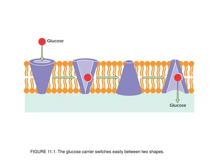
FIGURE 11.1. The glucose carrier switches easily between two shapes. UNFIGURE 11.1. .

E N D
FIGURE 11.1. The glucose carrier switches easily between two shapes.
FIGURE 11.2. (A) The monomeric oxygen-carrying protein myoglobin. Illustration: Irving Geis. Rights owned by Howard Hughes Medical Institute. Reproduction by permission only. (B) Oxygen binding of myoglobin (in red) and hemoglobin (in purple) as oxygen pressure increases.
FIGURE 11.3. A pH change that alters the charge on histidine will alter the balance of forces within a protein. At pH 6, the structure on the right will predominate.
FIGURE 11.4. Phosphorylation changes the charge pattern, and hence the balance of forces within the calcium ATPase, forcing a change of shape.
FIGURE 11.5. Catalysts act by reducing the activation energy (A) of a reaction.
FIGURE 11.6. Definition of v0, the initial velocity of a chemical reaction.
FIGURE 11.7. v0 measured in a number of reaction tubes (with [E] constant and always less than [S]) forms a hyperbolic curve when plotted as a function of substrate concentration.
FIGURE 11.8. Aminotransferases use a cofactor that participates in the reaction but ends up unchanged.
FIGURE 11.9. Phosphofructokinase is regulated by the binding of ATP or AMP at a regulatory site that is separate from the active site. Binding of ATP inhibits while binding of AMP activates.
FIGURE 11.10. Control of Cdk1 by cyclin B and by phosphorylation.