Download
1 / 13
140 likes | 372 Views
Cardiovascular Physiology. Lab #10. Path of Cardiac Excitation. Sinoatrial (SA) Node pacemaker of the heart Atrioventricular (AV) Node Delays conduction to ventricles Bundle of His conducts signal through interventricular septum Purkinje fibers

E N D
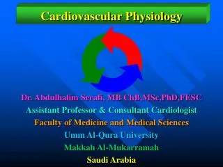
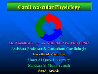
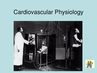
Cardiovascular Physiology Lab #10
Path of Cardiac Excitation • Sinoatrial (SA) Node • pacemaker of the heart • Atrioventricular (AV) Node • Delays conduction to ventricles • Bundle of His • conducts signal through interventricular septum • Purkinje fibers • conduct signal up lateral walls of ventricle Lecture Fig 13.19
Path of Cardiac Excitation • SA node cells produce APs • Atrial fibers activated • atrial contraction • APs excite AV node • delay (complete atrial contract) • APs of AV node travel down AV bundle to apex of heart • signal conducted to Purkinje fibers throughout ventricles • Myocardial fibers activated • ventricular contraction Lecture Fig 13.19
Electrocardiogram (ECG) • P wave • depolarization of atria just before contraction • QRS wave • depolar. of ventricles just before contraction • also atrial repolarization • T wave • repolarization of the ventricles Lecture Fig 13.21
Electrocardiogram (ECG) • P-R interval • Atrioventricular delay • R-T interval • Duration of ventricular systole • T-R interval • Duration of ventricular diastole P-R R-T T-R
ECG Exercises • Record ECGs before and after exercise • Measurements • Duration of a cardiac cycle (T-T) • Measurement of heart rate • Measurement of atrial systole and the A-V delay (P-R) • Measurement of ventricular systole (R-T) • Measurement of ventricular diastole (T-R)
Cardiac Cycle • contraction (systole) + relaxation (diastole) of ventricles • lasts 0.8 sec (based on 72 beats/min) Lecture Fig 13.12
Cardiac Cycle - Heart Sounds • “lub” = closing of the AV valves • “dub” = closing of the semilunar valves Lecture Fig 13.12
Auscultation • Listen for the heart sounds w/ stethoscope • Best heard in different positions Lecture Fig 13.14
Arterial Blood Pressure • Pressure blood exerts on arterial walls • Systolic blood pressure • pressure of blood in arteries during ventricular systole • Diastolic blood pressure • pressure of blood in arteries during ventricular diastole • Indicates blood flow to the body and work load of the heart Lecture Fig 14.15
Measure Blood Pressure • Sphygmomanometer • Apply cuff • Apply pressure to ~180 mmHg • Release pressure slowly • Auscultate brachial artery for sounds of Korotkoff Lecture Fig 14.29-14.30
Cardiovascular Fitness • Regular exercise • Increased stroke volume • Greater cardiac output • Can maintain exercise longer • Less increase in HR needed to meet blood flow demands • Activity of heart muscle itself is lower • Can recover from exercise more quickly • Can compensate for changes in blood flow due to positional changes more effectively.
Fitness Activity • Measure reclining and standing HRs • determine change in pulse rate and score • Calculate change in systolic BP as you go from a reclining position to a standing position • Perform exercise on stool, (3 seconds each cycle, 5x) record HR (15 sec x4) • measure pulse at 30, 60, 90 and 120 sec after completion (15 sec x 4) • record time for pulse to return to normal standing rate. • subtract normal HR from exercise HR • Tally up scores and see how fit you really are!