Download
1 / 34
420 likes | 799 Views
Neurons, synapses and signaling. The neuron – structure and function. Conducts long distance electrical signals and short distance chemical signals. Cell body – includes nucleus and other organelles Dendrites – highly branched extensions that receive signals from other neurons

E N D
The neuron – structure and function • Conducts long distance electrical signals and short distance chemical signals. • Cell body – includes nucleus and other organelles • Dendrites – highly branched extensions that receive signals from other neurons • Axon – extension that transmits signals to other cells, can be long, branched at end
Dendrites Stimulus Figure 37.2 Axon hillock Nucleus Cell body Presynaptic cell Axon Signal direction Synapse Synaptic terminals Synaptic terminals Postsynaptic cell Neurotransmitter
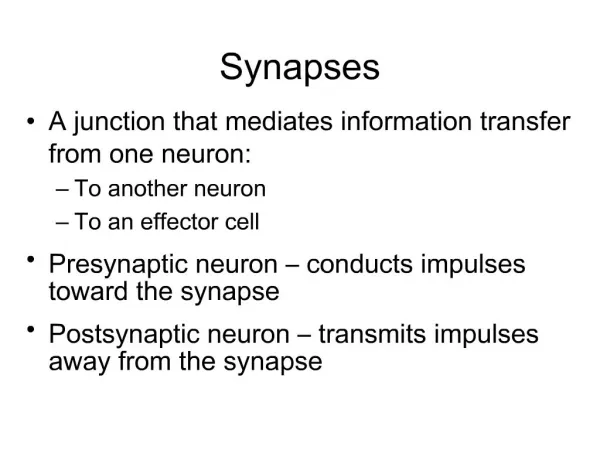
Synapse – junction between axon/dendrite • Neurotransmitters – chemical messengers, pass information from transmitting neuron to receiving cell • Glia cells – support cells in nervous system • Outnumber neurons • In brain • Nourish neurons, insulate axons, regulate the extracellular fluid around surrounding neurons
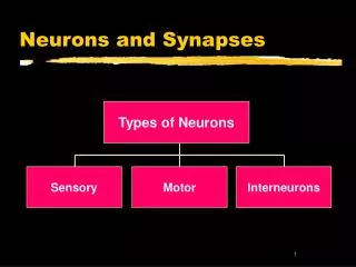
Information processing • Sensory neuron – interneuron – motor neuron • Central nervous system – brain and spinal cord • Peripheral nervous system – nerves • Autonomic nervous system – involuntary actions • Sympathetic – fight or flight • Parasympathetic – maintenance
Ion pumps and channels resting potential • Inside of a cell is negatively charged relative to outside • Membrane potential –charge difference between outside and inside of cell • attraction of opposite charges across the plasma membrane as source of potential energy • Resting potential – membrane potential for resting neuron
Resting potential • Potassium – K+ - greater inside of cell • Sodium – Na+ - greater outside of cell • Gradients are maintained by sodium potassium pump • S/P Pump – 3 K+ out for 2 Na+ in • Existence of a voltage difference in resting neuron • Ion channel – allows ions to move back and forth across the membrane and generates a membrane potential • Net flow of each ion across the membrane since neither K+ or Na+ is at equilibrium
Figure 37.6 Key OUTSIDE OF CELL Na K Sodium- potassium pump Potassium channel Sodium channel INSIDE OF CELL
Action Potentials - axons • Neuron responds to stimulus – gated ion channels react • Hyperpolarization – inside of membrane more negative due to opening K+ channel, which diffuse out, shifting membrane potential • Depolarization – reduction in the magnitude of the membrane potential, usually involves gated Na+ channels opening and diffusing into the cell • Action potential – massive change in membrane voltage
Figure 37.11 3 4 3 4 1 5 2 5 1 Key Na K Rising phase of the action potential Falling phase of the action potential 50 Action potential 0 Membrane potential (mV) 2 Threshold −50 1 Resting potential Depolarization −100 Time OUTSIDE OF CELL Sodium channel Potassium channel Undershoot INSIDE OF CELL Inactivation loop Resting state
Figure 37.12-3 1 2 3 Axon Plasma membrane Action potential Cytosol Na Action potential K Na K Action potential K Na K
Evolutionary adaptations of axon • Wider axon – allows for less resistance to the flow of currents • Invertebrates differ from vertebrates • Vertebrate axons have narrow diameters but do conduct action potentials at high speeds • Due to insulation – myelin sheath • Myelin sheaths in CNS – oligodendroglia in PNS – schwann cells
Figure 37.13 Node of Ranvier Layers of myelin Axon Schwann cell Schwann cell Nodes of Ranvier Axon Nucleus of Schwann cell Myelin sheath 0.1 m
Saltatory conduction • Myelinated axons have gaps – nodes of Ranvier • where voltge-gated Na+ channels are located • Action potentials occur at nodes and pass over myelinated sections – making conduction much faster
Figure 37.14 Schwann cell Depolarized region (node of Ranvier) Cell body Myelin sheath Axon
The synapse - communication • Electrical and chemical synapses • Most synapses are chemical synapses in the vertebrate brain • Release of neurotransmitters, held in vesicles, by the pre-synaptic neuron • Neurotransmitter diffuses across the synaptic cleft, to the post synaptic membrane, which activates a specific receptor
Figure 37.15 2 1 3 4 Presynaptic cell Postsynaptic cell Axon Synaptic vesicle containing neurotransmitter Synaptic cleft Postsynaptic membrane Presynaptic membrane K Ca2 Ligand-gated ion channels Voltage-gated Ca2 channel Na
Neurotransmitters • Acetylcholine – nervous system functions, muscle stimulation, memory formation, learning • Glutamate – AA – in invertebrates, at neuromuscular junction rather than acetylcholine • GABA – (gamma-aminobutyric acid) – inhibitory synapses, increase permeability to Cl-, Valium reduces anxiety through binding to a site on a GABA receptor • Norepinephrine - excitatory • Dopamine and serotonin – affect sleep, mood, attention and learning, Parkinsons, depression • Endorphins – decreasing pain perception
Evolution of the nervous system in the Animal Kingdom • Cnidaria – nerve net, contraction and expansion of gastrovascular cavity • Planarian – cephalization – eye spot, nerves, nerve cords, simple CNS • Insects – ganglia –clusters of neurons, brain, ventral nerve cord • Vertebrates – CNS – brain and spinal cord • PNS - nerves
Eyespot Brain Nerve cords Figure 38.2 Transverse nerve Nerve net (a) Hydra (cnidarian) (b) Planarian (flatworm) Brain Brain Spinal cord (dorsal nerve cord) Ventral nerve cord Sensory ganglia Segmental ganglia (d) Salamander (vertebrate) (c) Insect (arthropod)
Glia cells • Nourish, support and regulate the functioning of neurons • Astrocytes – hold blood vessels close, aid in nourishment • Oligodendroglia – make myelin sheath in CNS • Microglia – phagocytic,
CNS - PNS • Gray matter – consists mainly of cell bodies and dendrites • White matter – consists of myelinated axon bundles • Brain – consists of 100 billion neurons
Figure 38.4 Peripheral nervous system (PNS) Central nervous system (CNS) Brain Cranial nerves Spinal cord Ganglia outside CNS Spinal nerves
Figure 38.5 Central Nervous System (information processing) Peripheral Nervous System Afferent neurons Efferent neurons Autonomic nervous system Motor system Sensory receptors Control of skeletal muscle Enteric division Parasympathetic division Internal and external stimuli Sympathetic division Control of smooth muscles, cardiac muscles, glands
Figure 38.6b Brain structures in child and adult Embryonic brain regions Cerebrum (includes cerebral cortex, white matter, basal nuclei) Telencephalon Forebrain Diencephalon (thalamus, hypothalamus, epithalamus) Diencephalon Mesencephalon Midbrain (part of brainstem) Midbrain Pons (part of brainstem), cerebellum Metencephalon Hindbrain Myelencephalon Medulla oblongata (part of brainstem) Diencephalon Mesencephalon Cerebrum Metencephalon Midbrain Myelencephalon Diencephalon Hindbrain Midbrain Pons Medulla oblongata Spinal cord Telencephalon Forebrain Cerebellum Spinal cord Embryo at 5 weeks Child Embryo at 1 month
Brain region functions • Cerebrum – skeletal muscle contraction, center for learning, emotion, memory and perception • Cerebellum – coordinates movement and balance, learning and remembering motor skills. • Diencephalon – • thalmus– input center for sensory information • Hypothalmus- thermostat, biological clock • Regulates pituitary gland therefor regulates hunger and thirst, fight or flight, role in sexual and mating behaviors.
The brain stem • Midbrain – receives sensory information, coordinates visual reflexes • Pons and Medulla – 2 way conduction from spinal cord to brain • Helps to coordinate large scale body movements, control several automatic, homoestatic functions: breathing, heart and blood vessel activity, swallowing, vomiting and digestion.
Figure 38.6d Diencephalon Thalamus Pineal gland Brainstem Hypothalamus Midbrain Pituitary gland Pons Medulla oblongata Spinal cord
Emotions – Limbic system • Biological clock regulation – • Typically regulated by cycles of light and dark • Coordinated by a group of neurons n the hypothalmus in conjunction with sensory information from the eyes. • Brain reward system and drug addition • Drugs alter the transmission of signals in the synaptic pathway formed by neurons. • Mouse party Use imaging of the brain to understand the brain Positron emission tomagraphy (PET) Magnetic resonance imaging (MRI)
Figure 38.8 Thalamus Hypothalamus Olfactory bulb Hippocampus Amygdala
Cerebral Cortex • Controls • Language and speech – Broca’s area and Wernicke’s area • Both in left side of brain… Left side of brain is also more adept at math and logical operations Right side – recognition of faces and patterns, spatial relations and nonverbal thinking. Frontal lobe – decision making
Figure 38.11 Somatosensory cortex (sense of touch) Motor cortex (control of skeletal muscles) Parietal lobe Frontal lobe Sensory association cortex (integration of sensory information) Prefrontal cortex (decision making, planning) Visual association cortex (combining images and object recognition) Broca’s area (forming speech) Occipital lobe Temporal lobe Visual cortex (processing visual stimuli and pattern recognition) Auditory cortex (hearing) Cerebellum Wernicke’s area (comprehending language)
Evolution of cognition in Vertebrates • Perception and reasoning that constitute knowledge • Human evolution…larger cranial capacity • Hypothesis – evolution of a highly convoluted cerebral cortex • Primates, and cetaceans (whales and dolphins) • Birds – lack convoluted cortex but have organization of clustered neurons in top layer of brain, the pallium
Senses • Sensory receptor – sensory transduction - transmission – perception • Types of sensory receptors • Mechanoreceptors – pressure, touch, stretch, motion and sound • Electromagnetic - light, electricity and magnetism • Thermoreceptors – heat and cold • Pain – extreme pressure or temp • Chemoreceptors – solute concentration, smell, taste