Download
1 / 74
750 likes | 784 Views
This module covers musculoskeletal system components, injury types, assessment techniques, and treatment guidelines for EMS providers. Learn about fractures, dislocations, sprains, strains, and more. Gain practical skills in splinting, traction, and cervical collar application. Understand the significance of compartment and crush syndromes in emergency situations. Proper assessment and care can prevent complications in musculoskeletal injuries. Earn CE credits and enhance your emergency response capabilities.

E N D
Assessing and Treating Musculoskeletal Injuries May 2012 CE Condell Medical Center EMS System Site Code: 107200E -1212 Prepared by: Sharon Hopkins, RN, BSN, EMT-P Rev 6/14/12
Objectives Upon successful completion of this module, the EMS provider will be able to: • 1. Discuss components and function of the muscular and skeletal systems. • 2. Predict injuries based on the mechanism of injury. • 3. Differentiate between fractures, dislocations, sprains, and strains. • 4. Describe the six P’s evaluated during a musculoskeletal assessment. • 5. Explain the general guidelines for splinting. • 6. Describe signs and symptoms of compartment syndrome.
Objectives cont’d • 7. Describe complications of compartment syndrome. • 8. Describe complications of crush syndrome. • 9. Demonstrate proper measurement and placement of a cervical collar. • 10. Demonstrate proper application of the KED. • 11. Demonstrate proper application of the HARE traction (or similar traction based on your department). • 12. Demonstrate standing take down with the back board. • 13. Successfully complete the post quiz with a score of 80% or better.
Components - Musculoskeletal System • Composed of: • Bones (dense connective tissue) • Joints (place where bones meet) • Muscles (tissues or fibers) • Skeletal (voluntary), smooth (involuntary), cardiac • Cartilage (connective tissue) • Tendons (bands of connective tissue) • Ligaments (connective tissue)
Function - Musculoskeletal System • Provide the framework of the body • Support and protect internal organs • Allow movement of body parts or organs • Storage of salts and minerals • Production site of red blood cells
Bone Marrow • Highly vascular • Manufactures important blood components
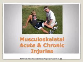
Musculoskeletal Injuries • Strain • Muscle injury from overstretching or overexertion of the muscle • Spain • Stretching or tearing of ligaments
Musculoskeletal Injuries • Dislocation • Disruption of a joint • Fracture • Any break in a bone • Simple = closed fracture • Compound = open fracture • Increased risk of contamination & infection • Most common bone injury
Cascade of Events • Fracture occurs Destruction of blood vessels in periosteum & bone and damage to surrounding vessels • Swelling of soft tissue • Formation of a clot in the area • Cell death at injury site due to disruption of blood flow • Intact surrounding cells divide & form a mass around fracture site • New bone is generated in weeks or months
Assessment Musculoskeletal Injuries • “5 P’s” of evaluation • Pain or tenderness? • Pallor – paleness or poor capillary refill? • Paresthesia – pins and needles sensation? • Pulses – diminished or absent? • Paralysis – inability to move?
Signs & Symptoms • Pain and tenderness • Usually localized • Deformity • Compare for symmetry • Grating or crepitus • Increases pain levels • Swelling • From bleeding at the site • Remove watches, rings as soon as possible • Document what you did with the personal effects
Signs & Symptoms cont’d • Bruising- leaking of blood vessels • Exposed bone ends • Open/comminuted fracture • Increases risk of infection • Bone infection could lead to amputation • Joints locked into place • Often seen with dislocations • Splint in position found
Signs & Symptoms cont’d • Nerve & blood vessel compromise • Evaluate distal CMS/SMV/PMS • Evaluated before and after splinting DOCUMENT CMS/SMV/PMS!!! Document ALL assessment results
Assessment PEARL • During assessment, determine mechanism of injury • If patient fell, ask “WHY” • If fall related to tripping/losing balance, you are just dealing with the orthopedic injuries • If patient experienced dizziness, lightheadedness, wooziness, syncope, near-syncope… • Consider a cardiac event until proven otherwise • Consider need for EKG monitoring • Perform the Cincinnati Stroke Scale
Care of the Injury • Standard Precautions observed • Perform baseline/initial assessment PEARL Musculoskeletal injuries are rarely ever life threatening Could be life threatening for bilateral femur fractures and pelvic fracture
Care of the Injury cont’d • Cover open wounds with sterile dressing • If life threatening situation, splint enroute if time • Note: Patients on backboard are essentially immobilized/splinted • If stable patient, can splint prior to transport
“RICE” • R – rest the injury (i.e.: splinting) • I – apply ice to wound • Never apply ice directly to the skin • Too damaging to the skin tissue and cells • C – apply compression to minimize swelling • Never pull tight on the ACE – will be too constrictive; let ACE unroll easily • E – elevate higher than the heart
Guidelines for Splinting • Must immobilize the joint above and joint below the injury • Minimizes movement which will decrease pain • Prevents additional soft tissue injury to nerves, arteries, veins, and muscle • Prevents a closed fracture from becoming an open fracture • Minimizes blood loss • Minimizes additional injuries to the site
Deformity • May make splinting difficult • Chance of compromise to nerves, arteries, and veins • Distal tissue may die due to compromised blood flow • May need to add extra padding • May need to be creative in choosing splinting material
When to Realign Deformed Extremities • Distal extremity cyanotic • Distal pulses cannot be palpated • When in doubt, call Medical control • For relatively short transport times, most injuries can and should be splinted in position found
Realigning an Injury • Goal: • Align joint to anatomical position • Splints applied in position of anatomical function • Position mimics a normal, relaxed pose for the extremity • Fingers slightly curved for hands
Realigning an Injury • General guidelines to follow if necessary: • 1 person grasps the distal extremity • 1 person places hands above & below injury • Apply gentle manual traction in the same direction as the long axis of the extremity • Stop if resistance is felt or bone ends may break thru the skin • Maintain gentle traction until splinting is accomplished
Splinting PEARLS • Can’t treat what you can’t see • Expose all injuries • Assess and document distal CMS/SMV/PMS before and after splinting • Consider need for padding around bony areas • If bone is protruding, do not push it back in • Cover with sterile gauze
Hazards of Splinting • Caring for extremity injuries prior to caring for life threatening injuries • Inappropriately staying on the scene to care for injured extremities prior to initiating transport • Improper or inadequate splinting • Too tight –circulation compromised • Too loose –movement allowed further injury
Potentially Fatal Orthopedic Injuries • Bilateral femur fracture • Typically results from excessive force • Consider the presence of additional injuries • Blood loss most likely with mid-shaft fractures • Can lose up to 2 units of blood (1000 ml) per femur fracture
Stages of Shock • Based on amount of blood loss • Stage 1 – up to 15% circulation volume • Average 500 – 750* ml (typical donation during blood drive) • Stage 2 – up to 15-25% circulation volume • Average 750 – 1250* ml • Stage 3 – up to 25-35% circulation volume • Average 1250 – 1750* ml • Stage 4 – up to >35% circulation volume *Averages calculated for a 70 kg person
Femur Fracture • Presentation • Extreme pain • A lot of muscle tissue surrounding the femur • Deformity • Swelling • Treatment • Traction splint • Best for mid shaft fractures
Traction Splinting • Relieves muscle spasm therefore reducing pain • Avoid if serious knee, tibial, or foot injuries • Avoid if any joint injury to hip or knee is suspected • Anterior hip fracture may look like a femur fracture • Head of femur often protrudes in inguinal area
Potentially Fatal Injury • Pelvic fracture • Frequently associated with extremity fractures • Usually result from MVC and falls from heights • Have high index of suspicion based on mechanism of injury • Can suffer from significant blood loss • Bones have rich supply of blood • Typically venous bleeding from disruption of bone surface
Pelvic Fractures • The most significant pelvic injury is open-book pelvic fracture • Symphysis is torn apart • Anterior pelvis opened like a book • Both sacroiliac joints usually disrupted
Pelvic Fracture • Assessment • Instability or pain when applying gentle posterior pressure on iliac crests or symphysis pubis during assessment • DO NOT ROCK PELVIS!!! • Could displace the fracture or disturb a hematoma • Up to 40% of patients also have abdominal injuries
Compartment Syndrome • Fascia is a non-stretching tough membrane that surrounds muscles and other structures in extremities • Multiple closed spaces created called compartments • Bleeding and swelling from trauma may create increased tissue pressure in the confined space
Compartment Syndrome cont’d • Increased pressure in confined space • Decreased blood flow • Hypoxia • Possible muscle, nerve, vessel impairment • May lead to cell death and amputation • Typically presents hours after initial insult • Surgical intervention required to relieve the pressures in compartment
Compartment Syndrome • Can occur with a patient with a casted extremity • Injured area continues to swell first few days • Casted area constricted and does not allow expansion of the swelling • Compartments become compromised • Have high index of suspicion for patient presenting with a cast • Pain level higher than expected usually the tip off
Signs and Symptoms Compartment Syndrome • Early • Pain out of proportion to injury • Paresthesia – pins & needles sensation • Late – 5 P’s • Pain • Pallor • Pulselessness • Paresthesia • Paralysis
Compartment Syndrome • Surgical intervention – fasciotomy • Will need to return to OR for closure at a later date
Compartment Syndrome • Risks of late diagnosis and intervention • Gangrene leading to need for amputation • Ischemic contractures and therefore loss of function • Rhabdomyolysis and acute renal failure • Syndrome caused by skeletal muscle injury • Leakage of large quantities of toxic intracellular contents into plasma • Basically, sludge of muscle protein attempting to be filtered thru kidneys is causing kidney damage
Crush Syndrome • Pressure on extremities during prolonged entrapment can disrupt blood flow • Typically 4 hours or longer of entrapment • Anaerobic metabolism in tissues occurs • Toxins produced & released from crushed tissues, muscles, and cells • Myoglobin - a muscle protein • Potassium • Phosphorus • Lactic acid – from anaerobic metabolism • Uric acid – from protein breakdown
Crush Syndrome cont’d • Patient at risk of cardiac dysrhythmia and severe kidney damage from toxins • Place patient on cardiac monitor • Watch for peaked T wave • Indication of excess potassium in vascular space • Increase IV fluid rate to keep kidneys hydrated and flushed
Hyperkalemia – High Potassium • Note peaked T wave (this is NOT ST elevation!!!) • Excess extracellular potassium is an irritant to the heart • Watch for dysrhythmias and potential arrest
Types of Splints • Rigid material • Air splint • Vacuum splint • Slings • HARE/Sager traction splint • Back board • Pillows
Cervical Collar PEARLS • Measure accurately for best fit • Improper fit causes greater risk of harm than it does good • Measure bottom of chin to top of shoulder • Eyes must be focused straight ahead
KED PEARLS • Helpful only when rapid extrication is not required • Maintain manual spinal motion restriction until fully secured • Carefully place the leg/thigh straps especially in the male population
HARE or Sager Traction PEARLS • Traction maintained manually until device in place and foot traction applied • Patients often experience instant relief of pain (from muscle spasms) once traction in place
Standing Backboard • Takes 3 persons to be safely performed • If you really need spinal motion restriction, doesn't make sense to have patient walk to cot and then lay down
Standing Backboard PEARL • Apply straps to finish securing the patient AFTER the patient is supine on the board • The patient will be manually held in place while the backboard is being lowered
Documentation • Assessment of injury by interview • Onset – what were you doing at the time? • Provocation/palliation – what makes the pain worse/better? • Quality – in your words, describe the pain • Radiation – does the pain radiate? • Severity – on a scale of 0-10, rate your pain • Time – what time did this happen?
Documentation cont’d • Observation of appearance • Blood loss present? • Deformity present? • Bruising present? • Assessment by palpation (CMS/SMV/PMS) • Pulses • Distal compared to proximal • Ability to wiggle distal extremities • Ability to differentiate area touched
Documentation cont’d • Consider the 6 P’s of extremity assessment • Pain • Pallor • Paralysis • Paresthesia • Pressure • Pulses