Download
1 / 26
260 likes | 361 Views
Tracking Volumetric Brain Deformation During Image Guided Neurosurgery. Alida Tei Graduate Student, Electrical Engineering and Computer Science Massachusetts Institute of Technology, Cambridge, MA, USA. Simon K. Warfield Florin Talos Aditya Bharatha Matthieu Ferrant. William M. Wells

E N D
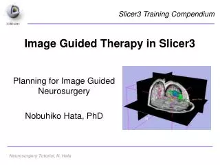
Tracking Volumetric Brain Deformation During Image Guided Neurosurgery Alida Tei Graduate Student, Electrical Engineering and Computer Science Massachusetts Institute of Technology, Cambridge, MA, USA
Simon K. Warfield Florin Talos Aditya Bharatha Matthieu Ferrant William M. Wells Ron Kikinis Ferenc A. Jolesz Peter McL. Black Acknowledgments URL: http://www.spl.harvard.edu
Presentation Outline • Challenges of neurosurgery • Tools for IGNS (Image Guided NeuroSurgery): • Image fusion • Nonrigid registration • Methodology (intraoperative): • Segmentation and rigid/affine registration • Nonrigid registration • Visualization • Atlas matching and results • Future plans
Challenges of Neurosurgery Removing as much tumor as possible without destroying healthy brain tissue • Healthy and diseased brain tissue have similar visual appearance • It is impossible to see critical structures underneath the brain surface
Tools for IGNS Images acquired intraoperatively help surgeon visualize lesions • Procedures carried out in operating rooms equipped with special imaging systems • MRI: high spatial resolution and soft tissue contrast
Signa SP (GE Medical Systems) Picture provided by R. Pergolizzi
Image Fusion for IGNS • Constraints of intraoperative imaging: time, magnetic field strength, only MRI • Before surgery: can use higher resolution MRI, different modalities (fMRI, SPECT, PET, MRA, diffusion) • Example: diffusion provides direct visual information on white matter tracts
Nonrigid Registration for IGNS During neurosurgery, nonrigid changes in anatomical position of brain structures (“brain shift”) • Reasons: CSF leakage, anaesthetic agents, hemorrhage, hyperventilation, retraction and resection, etc. • Consequence: preoperative data not as reliable during surgery
Brain Shift (1) Pictures provided by F. Talos
Brain Shift (2) Pictures provided by F. Talos
Methodology: Phase 1 • Intraoperative segmentation (brain and lateral ventricles): • automated multi-channel tissue classifier • binary curvature driven evolution algorithm • Rigid or affine registration of preoperative to intraoperative data
Methodology: Phase 2 Nonrigid registration (FEM): • Generation of unstructured surface and volumetric meshes • Surface matching: brain and ventricles iteratively deformed • Volumetric biomechanical solver, (brain as homogenous linearly elastic material) • Volumetric deformation fields applied to preoperative images
Active Surface Deformation Video provided by M. Ferrant
Methodology: Phase 3 Intraoperative visualization: critical structures extracted from preoperative data and visualized in operating room • Integrated system: intraoperative images displayed with surface models • Intraoperative navigation: virtual surgical instruments visualized in coordinate system of patient image acquisition
Atlas Matching during IGNS Brain structures mapped through image fusion of deformable volumetric atlas • Brain shift captured by FE model simulating brain elastic properties • Appropriate structures (according to tumor’s location) extracted from matched atlas and visualized • Clinical use made practical by real-time processing supported by HPC
Intraoperative Visualization Picture provided by F. Talos
Results • Brain deformation during surgery accurately calculated, critical brain structures tracked prospectively • Small registration error due to different anatomy of atlas and patient brain • Generic atlas, surrogate for preoperative data from different modalities (MRA, fMRI, diffusion tensor MRI, etc.)
Future Plans • Create patient-specific atlas • Incorporate diffusion tensor data • Expand biomechanical model (anisotropic inhomogeneous white matter; nonlinear, hyperviscoelastic framework) • Include physiological parameters measured during surgery