Download
1 / 22
220 likes | 404 Views
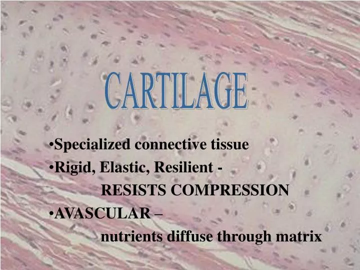
CARTILAGE. Specialized connective tissue Rigid, Elastic, Resilient - RESISTS COMPRESSION AVASCULAR – nutrients diffuse through matrix. Hundreds of Eyes Staring Back at YOU!. PERICHONDRIUM. Dense irregularly arranged connective tissue (type I collagen)

E N D
CARTILAGE Specialized connective tissue Rigid, Elastic, Resilient - RESISTS COMPRESSION AVASCULAR – nutrients diffuse through matrix
PERICHONDRIUM • Dense irregularly arranged connective tissue (type I collagen) • Ensheaths the cartilage • Houses the blood vessels that nourish chondrocytes
CHONDROBLAST • Progenitor of chondrocytes • Lines border between perichondrium and matrix • Secretes type II collagen and other ECM components • Chondroblasts build
CHONDROCYTE • Mature cartilage cell • Reside in a space called the lacuna • Clear areas = Golgi and lipid droplets
Chondrocytes completely fill their lacunae • RER and euchromatic nuclei • Synthetically active, secrete matrix N RER Cartilage matrix
MATRIX • Provides the rigidity, elasticity, & resilience • FIBERS • Collagenous and elastic • GROUND SUBSTANCE • Glycosaminoglycans (chondroitin sulfates, keratin sulfate, hyaluronic acid) • Proteoglycans: GAGs + core protein • Water • Basophilic • Territorial matrix - high [ ] of sulfated proteoglycans
CARTILAGE GROWTH • Appositional • Increasing in WIDTH; chondroblasts deposit matrix on surface of pre-existing cartilage • Interstitial • Increasing in LENGTH; chondrocytes divide and secrete matrix from w/in lacunae
TYPES OF CARTILAGE • HYALINE • ELASTIC • FIBROUS
HYALINE CARTILAGE • FUNCTION • Support tissue and organs • Model for bone development • MATRIX • Type II collagen (thin fibrils) • Chondroitin sulfate, keratin sulfate, hyaluronic acid • Water • LOCATION • Tracheal rings, nasal septum, larynx, articular surfaces of joints
ELASTIC CARTILAGE • FUNCTION • Support with flexibility • MATRIX • Normal components of hyaline matrix plus ELASTIC fibers • LOCATION • External ear, external auditory canal, epiglottis • STAINS • Elastic fibers stain BLACK with Weigert stain perichondrium
FIBROCARTILAGE Orcein van Giesen Elastic stain - fibrocartilage - reddish brown hyaline cartilage - yellow
FIBROCARTILAGE • FUNCTION • Support with great tensile strength • MATRIX • Type I collagen - Oriented parallel to stress plane • LOCATION • Intervertebral disks, pubic symphysis
FIBROCARTILAGE • Chondrocytes align between collagen fibers • Collagen fibers lie parallel to lines of stress
JOINT DEFINITIONS • SYNOVIAL CAVITY • Fluid filled space b/t 2 bones • SYNOVIAL FLUID • Water and GAGs; provides nutrients for cartilage • SYNOVIAL MEMBRANE • Continuous with the perichondrium
SYNOVIAL “MEMBRANE” Not a true membrane - Why? • Specialized secretory CT • Loose (areolar) CT • Formed by layers of collagen and fibroblasts • Highly vascular
No perichondrium • Joint capsule composed of DIACT • Why does the cartilage at the joint stain so acidophilic (i.e. pink)?
Question 1 Panel C is a low magnification micrograph of the tissue shown in Panel B. Provide the letter or letters (A and/or B) (or none) of the panel to which the following apply. normally calcified vascularized collagen type II cells capable of division matrix contains proteoglycans lacunae present A C B
C A Questions 2 and 3: 2. The three tissues shown have all of the following properties in common EXCEPT: a. They contain capillaries. b. They contain proteoglycans. c. They can increase in size by interstitial growth. d. They can increase in size by appositional growth. 3. Which tissue is the most highly specialized to resist compression? a. A b. B c. C B
Question 4 • 4. What cartilage nourishing tissue is missing at the interface shown? • Synovial fluid • Perichondrium • Synovial membrane • Chondroblasts