Download
1 / 65
910 likes | 3.22k Views
Corneal Dystrophies and Degenerations. Optometry 8570 Winter 2008. Definitions. Dystrophy: Primary tissue changes of genetic origin which occur in previously normal tissue Degeneration: Any tissue change occurring in previously normal tissue

E N D
Corneal Dystrophies and Degenerations Optometry 8570 Winter 2008
Definitions • Dystrophy: Primary tissue changes of genetic origin which occur in previously normal tissue • Degeneration: Any tissue change occurring in previously normal tissue • Congenital Anomaly: Gross morphological tissue alterations that manifest at birth
Bilateral Symmetrical Central Avascular Inherited Early onset (1-2 dec.) Slowly progressive Unrelated to systemic Dz Non-inflammatory Usually 1 corneal layer Unilateral Asymmetrical Peripheral Vascularized Not inherited Mid-life or later Progressive Often systemic Dz assoc. Variable inflammation Usually multiple corneal layers involved (Adapted from S.J. Grondalski, O.D.)Dystrophy Degeneration
Anterior Corneal Dystrophies • EBMD (map-dot-fingerprint dystrophy) • Reis-Buckler’s dystrophy • Meesmann’s dystrophy
Stromal Dystrophies • Lattice dystrophy • Granular dystrophy • Macular dystrophy • Central crystalline dystrophy (Schneider’s dystrophy) • Others
Endothelial Dystrophies • Fuch’s dystrophy • PPD • Congenital hereditary endothelial dystrophy (CHED)
Posterior Polymorphous Dystrophy (PPD) • Clinical Pearls: • One of the mesodermal dysgenesis syndromes • Autosomal dominant, presents in early life • Bilateral (vs. ICE syndrome) • M = F • Changes in Descemet’s result in epithelialization of the endothelium; proliferative tendencies • May appear as vesicles arranged in a linear or grouped pattern, usually as broad bands with irregular scalloped edges • May cause iridocorneal adhesions and/or decentered pupil (corectopia)
Congenital Hereditary Endothelial Dystrophy (CHED) • Clinical Pearls: • Bilateral corneal edema with normal corneal diameter, normal IOP, and no guttata • Autosomal recessive: present at birth, non-progressive, nystagmus present, pain and photophobia uncommon • Autosomal dominant: first seen in childhood, slowly progressive, pain/tearing/photophobia common
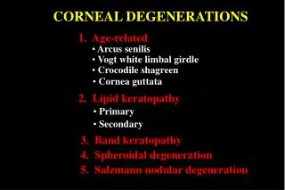
Degenerations • Depositions (corneal deposits of material that shouldn’t be there) • Keratoconus • Peripheral degenerations (Marginal thinning disorders)
Depositions • Arcus (lipid) • Band keratopathy (calcium) • Salzmann’s nodular degeneration • Spheroidal degeneration • Amyloid degeneration • Coat’s ring • Systemic meds
Keratoconus • Clinical Pearls: • Usually bilateral and asymmetric • Slowly progressive irregular astigmatism secondary to paracentral thinning and bulging of the cornea • Vogt’s striae: vertical tension lines in the posterior cornea • Irregular retinoscopic reflex • Irregular keratometry mires • May be associated with chronic eye rubbing (atopy) • Corneal hydrops (sudden corneal edema) results from rupture of Descemet’s, and may cause sudden reduced vision, pain, red eye, and photophobia
Keratoconus • Associated Etiologies: “AMANDA LEAH” • Atopy • Marfan’s syndrome • Addison’s Dz • Down’s syndrome • Amyloidosis • Leber’s congenital amaurosis • Ehler’s Danlos syndrome • Apert’s syndrome • Hypothyroidism