Download
1 / 1
20 likes | 412 Views
Cystic Adenomatoid Malformation of the lung. Lim Ri Kyung. Lee Eun Hey. Yun Sun Young. Choe Su Yearn Department of Obstetrics & Gynecology, The Catholic University of Korea Seoul. Mary's Hopital. Object

E N D
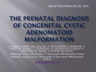
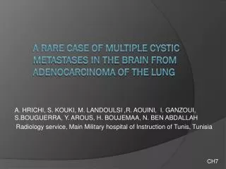
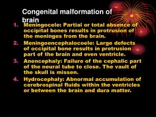
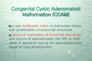
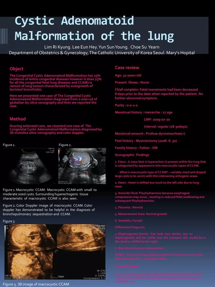
Cystic Adenomatoid Malformation of the lung Lim Ri Kyung. Lee Eun Hey. Yun Sun Young. Choe Su Yearn Department of Obstetrics & Gynecology, The Catholic University of Korea Seoul. Mary's Hopital Object The Congenital Cystic Adenomatoid Malformation has 25% incidence of entire congenital diseases however it does 75% for all the congenital fetal lung diseases and CCAMs is consist of lung tumors characterized by overgrowth of terminal bronchioles. Here we presented one case of The Congenital Cystic Adenomatoid Malformation diagnosed from a 20w+1d of gestation by ultra-sonography and then we reported the case. Method Duaring antenatal care, we resented one case of The Congenital Cystic Adenomatoid Malformation diagnosed by 3D mutislice ultra-sonography and color doppler. Case review Age: 40 years old Present illness : None. Chief complain: Fetal movements had been decreased 8 days prior to the date when reported by the patient. No further abnormal symptom. Parity : 0-0-1-0 Menstrual history : menarche : 17 age LMP: 2009-07-20 interval: regular (28-30days) Menstrual amount : Profuse dysmenorrhea(+) Past history : Myomectomy (2008. 8. 30) Family history : Father - DM Sonographic Findings 1. Fetus : A mass that is hyperechoic is present within the lung that is categorized by appearance into macrocystic types of CCAM. : What is macrocystic type of CCAM? – variably sized and shaped large cysts (2 to 10cm) with thin intervening echogenic areas. 2. Heart : Heart is shifted too much to the left side due to lung mass 3. Amniotic fluid: Ployhydramnios because esophageal compression may occur , resuting in reduced fetal swallowing and subsequent Ployhydramnios. 4. Placenta : Normal 5. Measurement Data: Normal growth 6. Genitalia: Female Differential Diagnosis 1. Diaphragmatic hernia – Can look very similar , but no diaphragmatic will be visible and the stomach will usually be in the chest or shifted to the right. 2. Bronchopulmonary sequestration Pitfalls : Acoustic enhancement posterior to the heart and create the impression of a microcystic mass. Conclusions It is very important to have early diagnosis by ultrasound as soon as possible for fetal anormaly because prenatal counseling and antenatal care. Figure 1. Figure 2. Figure 1. Macrocystic CCAM. Macrocystic CCAM with small to moderate sized cysts. Surrounding hyperechogenic tissue characteristic of macrocystic CCAM is also seen. Figure 2. Color Doppler image of macrocystic CCAM. Color doppler has demonstrated to be helpful in the diagnosis of bronchopulmonary sequestration and CCAM. Figure 3 Figure 3. 3D image of macrocystic CCAM