Download
1 / 83
830 likes | 1.03k Views
Vascular Problems, Stroke, Aneurysms, and HTN Crisis. By Diana Blum MSN MCC NURS 2140. Vascular Disorders. Common disorders in America: hypertension atherosclerosis arterial occlusive disease abdominal aortic aneurysms (AAA) deep vein thrombosis (DVT)

E N D
Vascular Problems, Stroke, Aneurysms, and HTN Crisis By Diana Blum MSN MCC NURS 2140
Vascular Disorders • Common disorders in America: • hypertension • atherosclerosis • arterial occlusive disease • abdominal aortic aneurysms (AAA) • deep vein thrombosis (DVT) • venous insufficiency
hormones • C reactive protein is a marker for cardiac inflammation • Increases mean: risk of damage • Homocysteine: protein that promotes coagulation by increasing factor 5 and factor 11 while depressing activation of protein C and increasing thrombus formation risk • Vitamin b6 and b12 and folate lowers homocysteine levels
Arterial diseases: • Arteriosclerosis (atherosclerosis) • Aneurysm formation • Arteriosclerosis obliterans • Raynaud’s phenomenon • Arterial embolism • Thromboangiitis obliterans • Diabetic arteriosclerotic disease • hypertension
Manifestations :ARTERIAL(50% occulsion before symptoms) • Ischemia (reduced oxygenation) • - leads to pain • Paresthesia (decreased sensation in • extremities = tingling/numbing) • Pain (in feet/leg muscles = burning, • throbbing, cramping) • -usually from exercise BUT also • with elevation of lower extremities
(continued): • Hallmark sign: Intermittent claudication (pain in • exercising muscles – usually in calf • - directly related to decreased • blood supply during activity & • recedes with rest • Temperature: (COLD) • Skin color changes: skin pale on • elevation but red dependent
(continued) • Reactive hyperemia: (reduced blood flow to extremity results in arteriolar dilation so when the blood supply is restored, the affected area becomes warm/red from congestion • Pulse changes: Peripheral diminished or absent
(continued) • Prolonged capillary refill: • - 3 seconds or more • Ulcers: • - open lesions on feet from diminished distal perfusion
Arteriosclerosis • -describes arterial disorders in which • degenerative changes result in • decreased blood flow • Atherosclerosis: • - most common form of arteriosclerosis, excessive accumulation of lipids
Major risk factors of arteriosclerosis: • Hypertension (MOST SIGNIFICANT) • Cigarette smoking (nicotine has DIRECT • vasoconstricting effect) • Elevated serum cholesterol (fat causes • obstructive plaques) • Obesity (increased work to heart) • Diabetes (hyperglycemia causes damage to vessel wall) • Other: increase age, inactivity, family hx
Most common affected areas from arteriosclerosis: • Heart: coronary arteries (angina, MI, • death) • Brain (transient ischemic attacks =TIAs • CVA, death) • Kidneys (renal arterial stenosis lead to • chronic renal failure) • Extremities (gangrene of digits & • intermittent claudication)
Pathophysiology of atherosclerosis • -inflammatory process, begins as fatty streaks that are deposited in the intima of the arterial wall • Genetics and environment play a factor in the progression • Elastic arteries: aorta, carotid, lg & med. sized muscular arteries (popliteals) most susceptible arteries. • Endothelial injury: may be initiated by smoking, hypertension, diabetes, hyperlipidemia,
Inflammatory cells(including macrophages) become attracted to the wall • Macrophages infiltrate wall and ingest lipid which turns them into foam cells • They then release biochemical substances that cause further damage and attract platelets which then causes clots to form
Ankle-brachial index of blood pressure:Used to diagnose peripheral vascular disease • -compares the blood pressure at ankle with that of the arm. • -normally these should be the same (with a ratio of 1) • -lesser number than 1 shows decreased blood pressure at the ankle compared to upper extremity = = which indicates peripheral vascular disease to lower extremities
SURGERY • Indications for fem-pop bypass: • diabetes • hypertension • vasculitis • collagen disease • Bueger’s disease • Also, Embolectomy (surgical removal)
MEDICAL MANAGEMENT • ANTIPLATELET THERAPY • Aspirin, ticlid, plavix, pletal, trental • Beta blockers • ARBs • Statins • Radiation therapy • Angioplasty with stents
Nursing Interventions • Monitor BP for difference between arms • Could be indicative of aortic coarctation • Narrowing of aorta lumen • Monitor for carotid bruits • Assess cap refill, pulses,skin
Acute arterial stenosis • Monitor for the 5 P’s • pain, sudden • pallor • pulselessness • paresthesias • paralysis
Acute peripheral arterial occlusion • may result from rupture and thrombosis of an atherosclerotic plaque, an embolus from the heart or thoracic or abdominal aorta, an aortic dissection, or acute compartment syndrome • Symptoms and signs are sudden
Buerger Disease • Autoimmune disease • Recurrent inflammation of small arteries and veins of the extremities resulting in thrombus formation and occlusion. • Unknown cause • Men 20-35 years old • All races • Link to heavy smoking/chewing tobacco • s/s: rubor (reddish blue) color to foot, no Pedal pulse, discolored legs when dangled, eventually gangrene sets in
Aneurysms of Central Arteries • Enlargement of artery to @ least 2X its normal • Aortic dissection • Medial & intimal layers separate • Risk Factors: • -hypertension • -cocaine use • - Marfan syndrome
Thoracic Aortic Aneurysm • 85% are caused by atherosclerosis • More frequent in men b/w 40-70 years old • Most common site for dissection • 1/3 of pts with this die from rupture
S/S • Asymptomatic • Pain is primary symptom—constant • Dyspnea • Cough • Hoarseness • Stridor • Aphonia (weakness or complete loss of voice) • Unequal pupils
Diagnostics • Chest x-ray • TEE • CT
Signs/symptoms of aortic dissection: • n/v, diaphoresis with pain • “tearing” pain • Sudden onset • not relieved with change of position • Dissection of ascending aorta: anterior CP with • radiation to neck, throat, jaw • Dissection of descending: interscapular back pain • radiation to lower back or abdomen
Treatment of hypertension for aortic dissection: • IV propranolol • Nitropresside drip after beta blocker ( nitropresside by itself causes tachycardia AND left vent. contractility that is why a beta-blocker should be given first, then start nitropresside drip) • Diagnosis: • CXR (but 10% normal) see medialstinal • widening • Contrast CT • MRI
GOAL: to keep blood pressure to lowest • possible but yet allows tissue perfusion • Per physican recommendations
Surgery for distal dissections: • Mortality in 1st 48 hrs if unrepaired proximal aortic dissections is 40% • Usually distal dissections treated medically unless: • rapid expansion • saccular formation • persistent pain • hemodynamic compromised • blood leakage • impending rupture
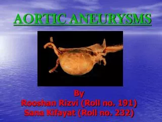
Abdominal Aortic Aneurysm (AAA) • 75% of all aneurysms Located between renal arteries & aortic bifurcation Symptoms from pressure exerted in surrounding structures. Many nonsymtomatic until ruptures Look for pulsating abdominal mass With rupture: hypovolemic shock & mortality around 90%
Nonsurgical management of AAA • Monitor growth: freq. CT scans • Antihypertensives • SURGICAL: • graft
Post-op nursing interventions for graft: • Vitals • Pulses distal to graft • Report: • changes in pulse • cool extremities distal to graft • white/blue to extremities distal to graft • severe pain • abd. distention • decreased UO
Post-op nursing intervention (continued)Post graft • Elevation of head to 45° or less • Renal function lab • Respiratory status • Paralytic ileus (NG tube) • Assess for dysrhythmias post thoracic
Venous diseases: • Venous thrombosis (thrombophlebitis) • known as DVT • Varicose veins • Venous stasis ulcers
Venous manifestations: • Pain: • - in feet/ leg muscles; aching/throbbing • - results from venous stasis & increases • as day progresses (esp with sitting • or standing) • Temperature changes: • - warm to touch since blood can enter • but cannot leave affected parts
Venous manifestations: • Skin color changes: reddened or • cyanotic • Edema: pooling of fluid results in edema • Venous stasis ulcers: skin breakdown • due to increased pressure from • chronic pooling of blood • Decreased mobility: may result from • the edema
DVT risk for pulmonary embolism • - legs • - seen post hip surgery, knee replacement pregnancy, ulcerative colitis, hrt failure, immobility
DVT : • Groin tenderness/pain • Unilateral sudden onset edema leg • Homan’s sign (appears in only 10% of pt • with DVT) • Ultrasonography
DVT interventions: • Rest (do NOT massage area) • Low-molecular weight heparin • Coumadin • TPA • ****Contraindications to anticoagulant therapy • Pt compliance, bleeding, aneurysms, trauma, alcohol, recent surgery, liver or kidney disease, hazard jobs, pregnancy
Nursing cares • Monitor for hemorrhage • Monitor PT/PTT • Heparin is therapeutic b/w 60-92 on ptt • Coumadin is therapeutic b/w 2-3 on PT/INR • Monitor for Thrombocytopenia • Monitor Platelets • s/s; purpura, bruising, hematomas • Provide bedrest • Ted Hose or ace wraps for prevention of DVT • SCDs for prevention of DVT • Pain meds
Hypertension • - excessive tension exerted on arterial walls which places pts at increased risk for target organ damage • -asymptomatic until complications develop • - elevation may be systolic or diastolic or both • - normal <120 mmHg systolic • <80 mmHg diastolic
S/S • Often none • Occipital headache more severe on rising • Lightheadedness • Epistaxis • Known as the ‘Silent Killer’