Download
1 / 10
100 likes | 302 Views
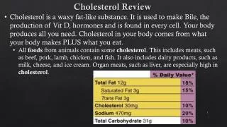
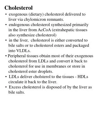
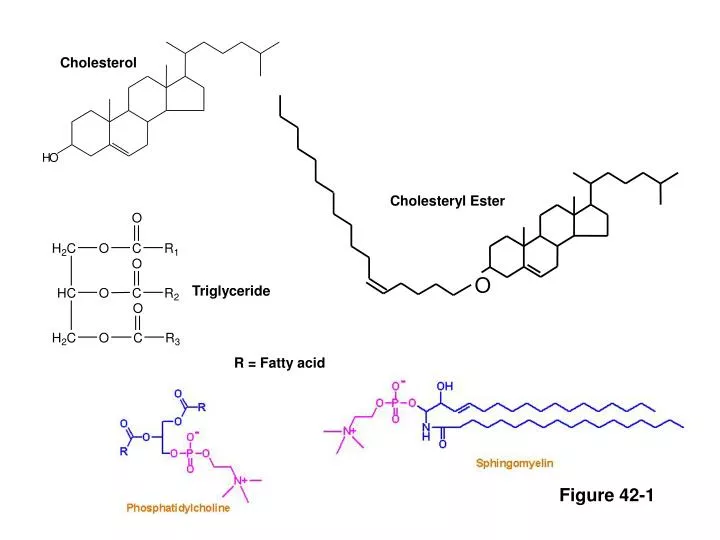
O. H. O. Cholesterol. Cholesteryl Ester. Triglyceride. R = Fatty acid. Figure 42- 1. Apolipoprotein. Phospholipid. Triglyceride. Cholesterol. Cholesteryl ester. Figure 42- 2. 0.95. Chylomicron. Chylomicron. VLDL. 1.006. IDL. Chylomicron remnant. 1.02. Density (g/mL). LDL.

E N D
O H O Cholesterol Cholesteryl Ester Triglyceride R = Fatty acid Figure 42-1
Apolipoprotein Phospholipid Triglyceride Cholesterol Cholesteryl ester Figure 42-2
0.95 Chylomicron Chylomicron VLDL 1.006 IDL Chylomicron remnant 1.02 Density (g/mL) LDL 1.06 HDL2 1.10 HDL3 1.20 5 10 20 40 60 80 1000 Diammeter (nm) Figure 42-3
FFA 2 3 Intestinal Pathway LPL Chylomicr. Remnant Chylomicron 1 10 Peripheral Cells Free Cholesterol ApoA-I, A-II ApoC-I, C-II, C-III Phospholipids Free cholesterol 7 6 Steroidogenic Cells HL, EL 8 LCAT HDL3 HDL2 Nascent HDL LDL 5 Triglycerides ApoA-I, A-II ApoC-I, C-II, C-III Phospholipids Free cholesterol 9 7 CETP PLTP Hepatic Pathway cholesteryl esters 2 3 6 HL VLDL LPL 4 IDL FFA Figure 42-4
IDL VLDL ApoB ApoE ApoB ApoE Endosome VLDL-R LRP ApoB LDL-R LDL Cholesterol HMG CoA Red ACAT Cholesteryl esters Fatty acids sER Lipoprotein assembly and secretion Bile acids VLDL Hepatic Cell Figure 42-5A
LCAT CETP PLTP Lipid-free apo AI Nascent HDL ABCA1 Endosome ApoB LDL-R LDL Cholesterol HMG CoA Red ACAT HDL3 sER Cholesteryl ester Stores Peripheral Cell Figure 42-5B
HDL SR-B1 Cholesterol Endosome ApoB LDL-R LDL Cholesterol HMG CoA Red ACAT sER Cholesteryl ester Stores Steroidogenic Cell Endothelial cell Hepatocyte Figure 42-5C
Fatty acids Insulin ASP Triglycerides HSL Fatty acids Adipocyte Figure 42-5D
Uptake of Oxidized Lipoproteins Efflux onto apo AI, apo E And HDL Apo Ai, Apo E Nascent HDL ABCA1 oxLDL ABCG1 SRA HDL Macrophage Figure 42-5E
Proportional reduction in event rate Reduction in LDL-C (mmol/L) Figure 42.6 From Baigent, C. et al., Lancet 2005;366(9493):1267-78