Download
1 / 34
510 likes | 1.12k Views
Nanofibers. Biophysics in the Guthold lab. Why study nanofibers. Have a physiological relevance Extracellular Matrix Blood Clots Gecko feet Spider scopula pads (setules) Interesting properties Surface area to volume ratio. Extracellular Matrix. Neural Interconnect and Cellular Matrix

E N D
Nanofibers Biophysics in the Guthold lab
Why study nanofibers • Have a physiological relevance • Extracellular Matrix • Blood Clots • Gecko feet • Spider scopula pads (setules) • Interesting properties • Surface area to volume ratio
Extracellular Matrix Neural Interconnect and Cellular Matrix Nerves and nerve bundles (yellow), extracellular supporting matrix (red), and ganglion cells (blue). Lust, University of Rochester University of Michigan Medical School Image
Blood Clot 50nm-200nm Colored SEM image of a whole clot Blue – Fibrin fibers Purple – platelet aggregation Red – red blood cells Credit: Yuri Veklich and John W. Weisel, University of Pennsylvania School of Medicine
Gecko Feet 200 – 500 nm spatula Van der waals interactions (electrostatic) Base on geometry not material Sitti and Fearing, Journal of Adhesion Science and Technology, 2003.
Our focus - 1Native Fibrin fibers (blood Clots) Heart attack Stroke DVT Hemorrhaging Embolism • Clots have been studied on the macroscopic level • No good model for blood clots • Gain a better understanding from the ground up • Model clot
The mechanical properties of native and gamma-crosslinking deficient fibrin fibers • Background and Motivation • Mechanical Properties of Native Fibers • Conclusions and Fibrin Fiber Model • Properties of Fibrin Fiber Branch Points
1.1. The Major Structural Component of a Blood Clot is a Network of Fibrin Fibers* Image: Yuri Veklich & John Weisel • Blood clots ‘perform’ the mechanical task of stemming the flow of blood Need to understand mechanical behavior of clot and its constituents. • One goal is to build a realistic model of a blood clot, based on the physical parameters of the fibers 1. • Another goal is to learn more about internal structure of fibers. • How does the clot perform, depending on numerous variables (mutations, environment, crosslinking, diseases, etc). *ignoring platelets for the time being 1 A. E. X. Brown et al, “Multiscale Mechanics of Fibrin Polymer: Gel Stretching with Protein Unfolding and Loss of Water” Science (2009) 325, 741-744
Fibrinogen A a b B Thrombin Dimensions of monomer: ~ 45 x 4.5 nm Crystal structure (chicken), Z. Yang, et al Biochemistry40, 12515-12523 (2001) Fibrinopeptides A & B Protofibril formation + Lateral aggregation and branching Further lateral aggregation SEM image (Roy Hantgan) of fibrin clot (plus platelets) 20-200 nm 10mm 1.2 Formation of Fibrin Fibers Fibrin (protofibrils) • Major interactions: • A:a interactions, D:D interface, B:b interactions • g-g crosslinks, a-a crosslinks
1.2. Properties of any Fibrous Networks Generally Depend on Three Parameters • The properties of the individual fibers • The properties of the branching points • The architecture of the network Chicken wire • If you want to design a model/structure out of fibers, it is important: • to know these three parameters, • and how they affect overall properties • Thus, we need to: • Determine fiber properties, branchpoint properties, and architecture • Determine and test macroscopic properties of structures • Iteratively compare experimental data, model data, improve model.
AFM tip Fibrin fiber 6 mm 8 mm substrate 12 mm x-y-z translator Microscope x-y stage Objective lens 1.3. Experimental set-up Instrumentation set-up: Side view of set-up: Top view of set-up: A B C Fibrin fiber Ridge AFM tip Linit L’ L’’ Ridge • Features: • Obtain images & movies of manipulation • Easy manipulation (nanoManipulator) • Obtain stress-strain curves of fiber deformation • Can apply larger force regime than in normal force measurement • Well-defined geometry
Breaking strength Maximum extension 1.4. Stress-Strain Curves of Single Fibrin Fibers Energy loss Elastic limit: Greatest strain a material can withstand without any measurable permanent strain remaining upon the complete release of the load. Extensibility: rupture strain = strain at which fiber ruptures. For elastic deformations: Y… Young’s modulus For viscous fluids: h … viscosity Polymers usually show viscous and elastic properties
A B Ridge Fibrin fiber AFM tip Groove e = 70% e = 0 C 20 mm e = 183% 2.1. Extensibility of Fibrin Fibers Uncrosslinked batroxobin E D Original length 332 ± 71 Partially crosslinked: emax = 330% Fully crosslinked: emax = 147% Thr + X 265 ± 83 50 mm Bat +X 226 ± 52 Thr - X 226 ± 72 Bat -X Lini 0% 100% 200% 300% 400% Extensibility DL/Linit W. Liu et al. “Fibrin Fibers have extraordinary extensibility and elasticity” Science (2006) 313, 634
2.2. Elasticity of Fibrin Fibers A B C Crosslinked thrombin e = 80% D E F 20µm e = 230% Partially crosslinked fibers: ~ 180%* Uncrosslinked fibrin fibers: ~ 60 – 120%* Fully crosslinked fibers: ~ 50 – 75 %* * Difficult to measure exactly G W. Liu et al Science (2006) 313, 634
2.3. Stress-Strain curves: Modulus and Strain Hardening • Fibrin fibers become stiffer at around 100% strain (sigmoidal change) • Uncrosslinked fibers stiffen by a factor of 3 (consistent) • Crosslinked fibers stiffen by a factor of 1.9 (inconsistent) • Slope is modulus (stiffness): • Uncrosslinked: 4 MPa (initial); 12 Mpa (high strain) • Crosslinked: 8 Mpa (initial); 15 Mpa (high strain) W. Liu et al. JTH (2010) “The mechanical Properties of Single Fibrin Fibers” 8, 1030-1036 Collet JP et al. PNAS (2005) 102, 9133-7
2.4. Radius dependence of Modulus (stiffness) • Fibrin Fibers become stiffer with decreasing radius • Thin fibrin fibers are denser than thicker fibers W. Liu et al. JTH (2010) “The mechanical Properties of Single Fibrin Fibers” 8, 1030-1036
2.5. Energy loss in fibrin fibers • Inscribed are in stress-strain curve corresponds to energy loss in pull cycle. • Uncrosslinked: little energy loss at low strains, 70% energy loss at high strains. • Crosslinked: higher energy loss at low strains, 70% energy loss at high strains. • Again, sigmoidal (two step) shape. W. Liu et al. JTH (2010) “The mechanical Properties of Single Fibrin Fibers” 8, 1030-1036
2.6. Incremental Stress-strain curves: Elastic and viscous components • Incremental stress-strain curves can be used to separate elastic and viscous (time-dependent) parts of modulus • Values see summary table W. Liu et al. JTH (2010) “The mechanical Properties of Single Fibrin Fibers” 8, 1030-1036
2.7. Incremental Stress-strain curves: Relaxation times t1 = 2 s t2 = 50 s • Two relaxation rates • Relaxation rates are: • independent of strain • independent of cross-linking
2.9. Summary Table, Mechanical properties of fibrin fibers, Kelvin Model, and Partially Crosslinked Fibers W. Liu et al. JTH (2010) 8, 1030-1036
3.1 Conclusions from Single, Native Fibrin Fiber Stress-Strain Measurements • We determined extensibility, elasticity, energetic behavior, total and relaxed elastic modulus (stiffness), stress relaxation behavior. • Despite nearly crystalline structure of fibrin fiber, fibers have large elasticities and extensibilities. • Monomer must be able to extend while keeping interactions intact • Crosslinking increases stiffness, reduces extensibility, little effect on relaxation times. • Two relaxation rates, sigmoidal strain hardening (two step), sigmoidal energy loss (two step).
3.2 Molecular Model Brown AEX et al. Biophys. J. (2007) 92, L30-41 Primalov PL et al. J. Mol. Biol. (1982) 159m 665-83. Collet JP et al. Blood (2005), 106, 3824-30 Falvo MR et al. JTH (2008), 6, 1991-3 Litvinov RI et al. Biochemistry (2007) 46, 9133-42
3.3 Molecular Model, main features • Assumes A:a interaction and D:D interface stay intact. • 1. Mechanism: Elastic interactions of alpha C-terminal domain, even across protofibrils. • 2. Mechanism: Alpha helical to beta strand transition, as suggested from single molecule experiments. • 3. Mechanism: Partial unfolding of gamma domain. Energy to stretch fibrin monomer is similar to thermal melting energy of gamma domain.
4.1 Properties of Fibrin Fiber Branchpoints • Joints are much more stable than we expected, fibers could be stretched to over 2.3 times their length, before joints broke (details, next slide). • In about 100 experiments we never saw the fiber fully unzip. • We often saw triangular architecture that may prevent unzipping, (perhaps originating from ‘trimolecular’ or tetramolecular junctions’1). • Slight helical structure of protofibrils may also stabilize joints. 1 Mosesson, MW et al. Ann NY Acad Sci (2001) 936, 11-30; Mosesson MW, Blood (1993) 82, 1517-1521; Mosesson et al. Proc Natl Acad Sci U S A. (1989) 86, 1113-1117; Ryan EA, et al. Biophysical Journal. (1999);77, 2813-2826.
4.2 Properties of Fibrin Fiber Branchpoints • Crosslinked and uncrosslinked joints ruptured at strains of 132% amd 146%. • Crosslinked fibers rupture 40% at the joint, 60% along the fiber. • Uncrosslinked fibers rupture 70% at the joints, 30% along the fiber. • Crosslinking stabilizes joints (makes fibers less extensible). • Overall joints are stabalizes by protofibril twisting, triangular architecture and crosslinking.
Our focus - 2Nanofibers for tissue engineering • ECM is composed of nanofibers • Regenerative medicine -> Tissue engineering • cell differentiation and proliferation has been related to scaffold mechanical properties • WFIRM – bladder from collagen and PGA • One branch with exceeding research and promise is electrospinning of biological polymers -> to form scaffolds
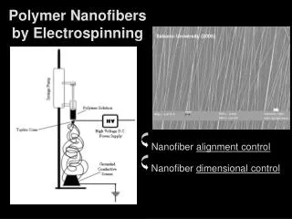
Working distance 1mm Post Taylor cone stream – MIT 2005, J.H. Yu, S.V. Fridrikh Electrospinning • Requirements for electrospinning • - High concentration polymer solution • - Volatile solvent, for example (HFP) • - Large voltage source • - Syringe and syringe pump • Candidate for tissue engineering scaffolds (Collagen and Fibrinogen) • - Biocompatible • - Support cell proliferation • Individual fiber properties important for design and engineering of structures with certain mechanical properties • By changing the polymer you can change the properties of the material Cell differentiation and proliferation have been related to mechanical matrix properties 25 μm
Stress-Strain curve of electrospun collagen Electrospun Collagen E A B C D Permanent deformation at all visible strains Significant strain softening Viscoelastic behavior such as stress relaxation Loses integrity in buffer unless crosslinked Large energy loss per cycle at strains over 12% Modulus dependence on radius
Electrospun Fibrinogen Rehydrated after spinning Strain softening (average total modulus 16 MPa, elastic modulus 6.7 MPa) Extensibility 133% Stress relaxation Relaxation fit with both double exponential and stretched exponential (convention) Modulus decreases with increasing radius
Electrospun fibrinogen and collagen • Collagen is soluble in buffer unless crosslinked • Fibrinogen and Collagen modulus decreases with increasing radius • Fibrinogen is more extensible • Collagen shows significant deformation at low strains • Both work well for cell seeding Current Work • Other polymers (PCL) • Combination spinning • Orientation • Layering
Electrospinning in Tissue EngineeringHeart Valve • Match Mechanical Properties of the tissue • Biodegradable • Cell adhesion • Shape • Function (three leaflets, prevent reverse flow)
Bioreactor Future Final Step • Seed Cells • Durability • Strong enough to be handled by physician during surgery; compliant enough to pump blood and degrade as cells produce their own ECM