Download
1 / 11
0 likes | 11 Views
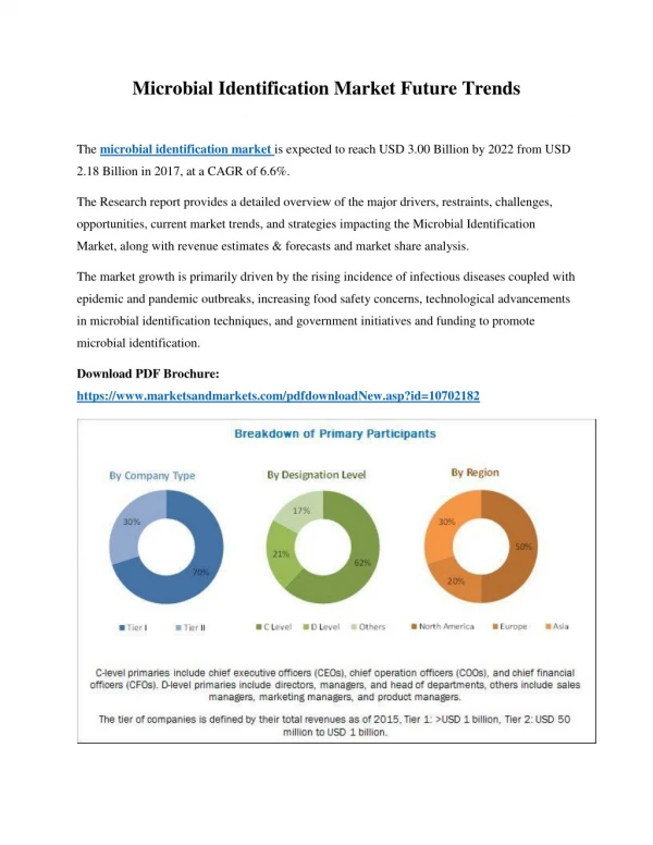
Water contamination is one of the major causes of water borne diseases worldwide. In Kenya, approximately 43% of people lack access to potable water due to human contamination. River Kuywa water is currently experiencing contamination due to human activities. Its water is widely used for domestic, agricultural, industrial and recreational purposes. This study aimed at characterizing bacteria and fungi in river Kuywa water. Water samples were randomly collected from four sites of the river: site A (Matisi), site B (Ngwelo), site C (Nzoia water pump) and site D (Chalicha),

E N D
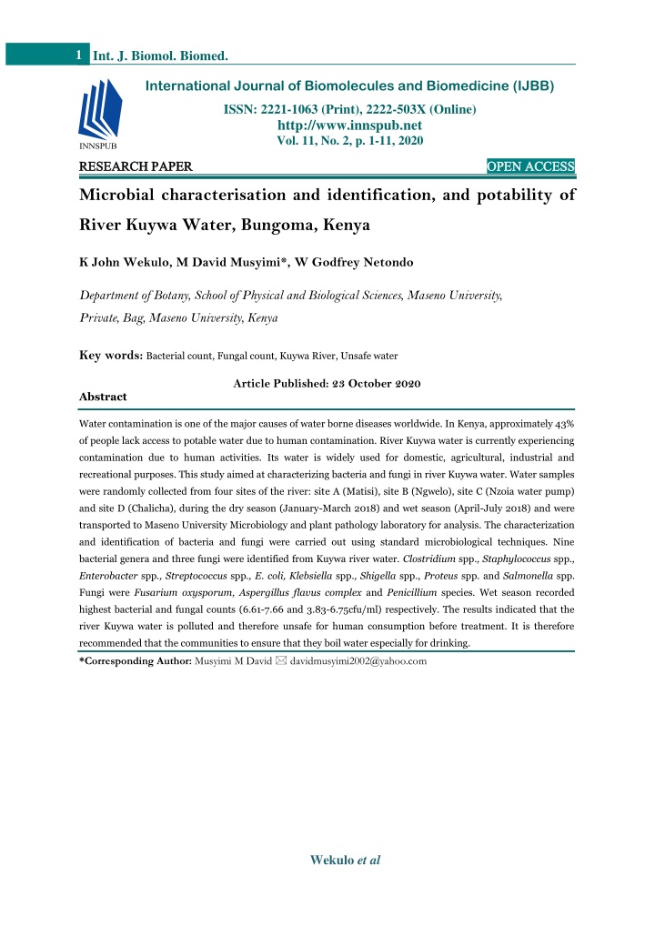
1 Int. J. Biomol. Biomed. International Journal of Biomolecules and Biomedicine (IJBB) ISSN: 2221-1063 (Print), 2222-503X (Online) http://www.innspub.net Vol. 11, No. 2, p. 1-11, 2020 RESEARCH PAPER RESEARCH PAPER OPEN ACCESS OPEN ACCESS Microbial characterisation and identification, and potability of River Kuywa Water, Bungoma, Kenya K John Wekulo, M David Musyimi*, W Godfrey Netondo Department of Botany, School of Physical and Biological Sciences, Maseno University, Private, Bag, Maseno University, Kenya Key words:Bacterial count, Fungal count, Kuywa River, Unsafe water Article Published: 23 October 2020 Abstract Water contamination is one of the major causes of water borne diseases worldwide. In Kenya, approximately 43% of people lack access to potable water due to human contamination. River Kuywa water is currently experiencing contamination due to human activities. Its water is widely used for domestic, agricultural, industrial and recreational purposes. This study aimed at characterizing bacteria and fungi in river Kuywa water. Water samples were randomly collected from four sites of the river: site A (Matisi), site B (Ngwelo), site C (Nzoia water pump) and site D (Chalicha), during the dry season (January-March 2018) and wet season (April-July 2018) and were transported to Maseno University Microbiology and plant pathology laboratory for analysis. The characterization and identification of bacteria and fungi were carried out using standard microbiological techniques. Nine bacterial genera and three fungi were identified from Kuywa river water. Clostridium spp., Staphylococcus spp., Enterobacter spp., Streptococcus spp., E. coli, Klebsiella spp., Shigella spp., Proteus spp. and Salmonella spp. Fungi were Fusarium oxysporum, Aspergillus flavus complex and Penicillium species. Wet season recorded highest bacterial and fungal counts (6.61-7.66 and 3.83-6.75cfu/ml) respectively. The results indicated that the river Kuywa water is polluted and therefore unsafe for human consumption before treatment. It is therefore recommended that the communities to ensure that they boil water especially for drinking. *Corresponding Author: Musyimi M David davidmusyimi2002@yahoo.com Wekulo et al
2 Int. J. Biomol. Biomed. Introduction Water bodies are areas of intense microbial activity Surface and underground water are major sources of and microbial interaction as nutrients and levels of fresh water but surface water is vulnerable to physicochemical parameters in water bodies are the contamination as contaminants can easily flow into it main source of food and driving forces for their (Harikumar et al., 2017). Water resources particularly proliferation and activities (Walker et al., 2014). The rivers in the world are degraded by discharge of microbial count in river water differs quantitatively untreated sewage and untreated industrial wastes. and qualitatively depending on level of contamination Free suspended bacteria in the water and bacteria (Verani et al., 2019). There is very limited, or no associated with the suspended materials are quoted published information, on river water quality amongst pollutants (Noble et al., 1997). assessment in Bungoma region yet disturbances from urban and agricultural development contribute to an Many people lack access to potable water and about overall decrease the biological integrity of a river. 22% of the world’s population per year are affected by There are no previous studies that have been water borne diseases (Akubuenyi et al., 2013; Karamage undertaken on characterization and identification of et al., 2016). Miime et al. (2011) reported that worldwide microbes in river Kuywa water. This study aimed at approximately 1.7 million deaths recorded annually are determining the the safety of river Kuywa water for attributed to unsafe water supplies. Contaminated water human consumption. serves as a medium of transmitting dangerous pathogens into humans, animals and plants and about Materials and methods 80% of human diseases are caused by water (Akubuenyi Study Site et al., 2013; Chen et al., 2017). Microbiological The study was done on river Kuywa in Bungoma contamination with faecal bacteria subsequent to County. The river source is Mt. Elgon and it flows anthropogenic activity is considered a crucial issue southwards through Bungoma County. It is a throughout the rivers in Kenya. tributary of river Nzoia, relatively wide, deep and it is lotic throughout the year. The river lies between In Kenya, approximately 17 million people (43%), lack latitude 34o00’ E and 35o00” E and longitude 0o 47' access to potable water, due to contamination of 24'' N and 0o 43' 40'' N and covers an area of about water sources. The main problems that affect water 110Km2 long(Wasike, 2015). The study area receives quality of the river are the high pollution following annual rainfall of 1500mm with above 850 mm in wet different human activities and population explosion. and less than 170 mm during dry season. Water sources in Kenya particularly rivers are Sample Collection becoming contaminated by both point and nonpoint Water samples were collected from four sites; Matisi sources attributed to human activities (Aywa, 2017). (Site A), Ngwelo (Site B), Nzoia water pump (Site C) River Kuywa flows southwards through Bungoma and Chalicha (Site D) (table 1) using clean sterilized County (Wasike, 2015). Its water is widely used for 250ml bottles from 20 to 30cm depth (to avoid domestic, agricultural, industrial and recreational floating materials) according to Mgbemena et al. purposes (Omwoma, 2011). This water body is feared (2012). The bottles were carefully closed and to be contaminated microbiologically due to effluents transported on ice and stored at 4⁰C in a refrigerator from coffee and sugar factories, domestic wastes, until the analysis of microbiological parameters. livestock excrements and agricultural activities. These Water samples were collected in triplicates once per may alter the microbiological parameters which season from the four different sites chosen based on eventually affects water quality of the river. More accessibility in terms of plants and animals, slope than 40,000 cases of typhoid and allergy in the region angle, human activities and health problems are reported annually (Wasike, 2015) which could be reported in the regions during the dry season attributed to water contamination. (January-March) and wet season (April-July) in Wekulo et al
3 Int. J. Biomol. Biomed. 2018 respectively. The sampling distance was about Culturing and determination of total bacteria count five kilometers between the sites. Serial dilution of water samples was carried out using distilled water to a dilution factor 105 and 0.1ml Table 1. Human activities around the sampling sites. aliquot from each dilution was well labelled and used for total plate count. One mililitre aliquot was spread Sampling sites Geographical position Characteristic on nutrient agar (NA) by pour plate technique and Latitudes Longitudes incubated for 24 hours at 37oC. Total bacteria count Bathing, Clothes washing, swimming fishing, livestock, agricultural activities Agricultural activities, waste disposal in water and swimming. Agricultural, effluents released from sugar factory Matisi (Site A) 00o37.186’N 034o42.094’E was done by counting visible and distinct colonies on the media using digital colony counter model Ngwelo (Site B) 00o34.412’N 034o40.825’E SUNTEX colony counter 570 and expressed as colony forming unit (cfu/ml). Nzoia water pump (Site C) 00o35.720’N 034o41.094’E Isolation of pure fungi colonies Twenty four water samples were subjected to culture Chalicha (Site D) 00o35.708’N 034o41.080’E technique using streak plate and spread plate techniques. A sterile wire loop was used to collect a loop full of each of undiluted water sample and Isolation of Pure Bacterial Culture inoculated on the surface of Potato dextrose agar. The The culture techniques were adopted from Mgbemena inoculated culture were sub-cultured on fresh media et al. (2012). Water culture samples were prepared by Potato dextrose agar plates to obtain pure cultures streak and spread plate techniques. A sterile wire loop which were used to further study their morphological was used to collect a loop full of each of undiluted characteristics. The pure culture isolates were sub- water sample and inoculated on the surface of cultured in Potato dextrose agar and incubated at room nutrient agar, MacConkey agar and Salmonella temperature for 3-7 days for fungal enumeration. Shigella agar, respectively. The inoculated culture Identification of fungal isolates were sub-cultured on fresh nutrient agar, MacConkey Identification of fungal isolates was done according to agar and Salmonella Shigella agar plates to obtain Oliveira et al. (2016). Visual observation of sub- pure cultures which were used to further study their cultured colonies characteristics (colour, shape, morphological and biochemical characteristics. The margin, elevation and presence of aerial mycelium) pure culture isolates were sub-cultured in nutrient was done. A phase contrast microscope was used to agar and incubated at 37oC for 24 hours for bacterial pre identify colonies of fungi which were emulsified enumeration. The samples were processed and on to glass slides using wet mount technique. analyzed to determine heterotrophic and coliforms Distilled water and lactophenol cotton blue staining using nutrient agar, MacConkey agar and Salmonella (LPCB) were utilized as mountants. The mycelium Shigella media. was teased (picked) out with the needles and covered with clean cover slip carefully avoiding air bubbles Morphological identification of bacteria isolates and observed under the microscope for shape, from water samples from river Kuywa conidia, conidiophore, arrangement of spores and The morphological characteristics of bacterial septation. The slide was mounted and observed under colonies such as shape, surface appearance, colour, magnification x400. Identity was confirmed with the margin, opacity and elevations were studied following help of mycology manual version 1.1. the procedures of Bergey’s manual of determinative bacteriology. The cell morphology was identified by Culturing and Determination of total Fungi Count placing pure bacterial colonies on the glass slide and Serial dilution of water samples was carried out using observed under light microscope for cell shape. distilled water to a dilution factor of 105. 0.1ml aliquot Wekulo et al
4 Int. J. Biomol. Biomed. from each dilution was labelled and used for total Citrate Test plate count. Samples of fungi were cultured on potato This was employed in determining the ability of bacteria to utilize sodium citrate as its only carbon dextrose agar (PDA). 0.1ml aliquot was spread on and energy source. The inoculated medium was potato dextrose agar by pour plate technique and incubated for 48 to 72 hours to allow complete incubated for 72 hours at 22oC. Fungi colonies were utilization of Simmon’s citrate medium by counted on the 3rd day using colour maker to spot on microorganisms. The colour of the medium indicated the lid. After 7 days, fungi colony were counted using the result. If the colour of media changed from green a different colour maker to spot only the colony that to blue then the bacteria was citrate positive while if grew on the plate and sum up the total colony number the media retained the green colour after incubation counted on 3rd and 7th day. period it indicated citrate negative bacteria. Methyl Red Test Gram Staining Technique A glass slide smear was prepared by placing deionized Tubes of MR-VP broth were inoculated by fresh pure water drop on the slide then microorganisms culture obtained from pure colonies. The inoculated obtained from the pure cultured colonies were MR-VP broth were incubated for 48-72 hours to allow aseptically transferred to the water drop using a microorganisms to fully utilize the supplied glucose, peptone and phosphate as explained by Behera (2012) sterilized wire loop and were spread with inoculating buffer at 37oC after which, one milliliter (1ml) of the loop. The smear was air dried; crystal violet was initial 30ml was transferred into test tubes. Three added to the smear then gently washed with tap drops of methyl red were added to them. Two test water. Iodine solution which is a mordant was added tubes were used per isolate, one tube was the control and allowed to stand for 2 minutes. Smear was then containing MR-VP broth inoculated with pure culture decolourized by using 95% ethanol, washed with tap and methyl red was not added after incubation water, counterstained with safranin for 45 seconds period. Another tube after incubation period, methyl and then washed with tap water. The smear was then red drops was added. Change in colour to red dried using filter paper and examined under oil indicated positive results while change in colour to immersion for gram stain. yellow indicated negative results. Voges Proskauer Test Indole Test A tube of MR-VP broth was inoculated with a pure This determined the ability of bacteria to split amino culture of the test organism and incubated for 24 acid tryptophan to form compound indole. One hours at 35oC. Oneml of broth was transferred to percent tryptophan 10ml broth was taken in test clean test tubes; 0.6ml of 5% alpha naphthol was tubes and inoculated by fresh pure culture obtained added, followed by 0.2ml of 40% KOH. Test tubes from pure colonies. After 48 hours of incubation were gently shaken to expose the medium to period at 37oC, one militre (1ml) of chloroform was atmospheric oxygen and then allowed to remain added to the broth. The test tubes were shaken gently. undisturbed for 15 minutes. Two test tubes were used Five drops of Kovács reagent was added directly to per isolate. Control test tubes contained MR-VP and pure culture obtained from pure colonies. Formation the tubes. These were also shaken gently and allowed of red colour indicated positive bacteria while brown to stand for twenty (20) minutes. Two test tubes were or yellow colour indicated negative bacteria. used per isolate with one being a control. Control test Carbohydrate Fermentation Test tube contained one percent tryptophan broth and inoculated by fresh pure culture obtained from pure The test was performed by inoculating 0.2ml of colonies. Formations of red colouration at the top nutrient agar, MacConkey agar and Salmonella layer indicated positive while yellow colouration Shigella agar culture of the isolated organisms into indicated negative results, respectively. the tubes containing 4% basic sugars; dextrose, Wekulo et al
5 Int. J. Biomol. Biomed. morphologically different based on colony size, shape, maltose, lactose and sucrose and incubated for 24 hours at 37oC. Acid production was indicated by the surface, margin, colour, elevation and opacity and cell colour change from red to yellow which showed shape (Table 2). Isolate A had circular shape, smooth bacteria fermenting the carbohydrate. Gas production surface, irregular margins, yellow colony colour, was noted by the presence of bubbles in inverted opaque and rods shaped. Isolate B was circular, rough (Durham) tubes due to fermentation. surface, irregular margins, white colony colour, translucent and cocci formed in chains. Isolate C had Catalase test irregular shape, mucoid surface, irregular margins, Catalase test was done to determine aerobic and cream in colour, opaque and rods shaped. Isolate D anaerobic bacteria and it was important in had a smooth surface, entire margin, orange colony differentiating morphologically similar Enterococcus colour, opaque and cocci formed separately. Isolate E Staphylococcus (catalase positive) and Streptococcus was circular, mucoid surface, entire margins, bright spp (catalase negative). Threeml of catalase reagent pink colony colour, raised, opaque and rods shaped. (3% H2O2) was put on a glass slide. Single colony Isolate F was circular, mucoid surface, entire margins, from the pure culture of bacteria from each sampled pale yellow, flat, translucent and cocci formed in site was scooped with a glass rod and submerged in chain. Isolate G was circular in shape, had smooth the reagent and observed for bubble formation which surface, entire margins, colourless colony colour, indicated positive test while absence of bubbles raised, opaque and rods. Isolate H had irregular formation indicated negative results. shape, smooth surface, filamentous, brick red colony colour, raised, opaque and rods.Isolate I had mucoid Oxidase test surface, entire margins, black colony colour, raised, The test was used to identify bacteria that produce opaque and rods shaped. cytochrome c oxidase, an enzyme of the bacterial electron transport chain. A filter paper was soaked in Gram staining 1% kovacs oxidase reagents and dried. A single colony The bacterial isolates A, B and D remained purple from pure culture of bacterial was rubbed into paper after decolourizing with alcohol confirming the using a wire loop. The colour change was timed using isolates as Gram positive while bacterial isolates C, D, a stop watch whereby, if colour changed to dark E, F, H and I were pink after decolourizing with purple within less than 10 seconds it indicated alcohol. This was an indication of Gram negative positive and if colour took more than four minutes to bacteria (Table 3). change it indicated negative results. Indole test Data Analysis Indole test of bacteria isolates A, DandE showed a Data on bacterial count and fungal count were development of a red ring at top layer of the medium subjected to Analysis of variance (ANOVA) using (Table 3) respectively. This was an indication of (SAS) version 9.1. Descriptive statistics including the indole positive bacteria. On the other hand, bacterial mean, standard deviation and standard error were isolates B, C, F, H, G and Iafter incubation period calculated at p = 0.05 confidence interval. Means that and on addition of Kovacs reagent formed a yellow were considered significantly different at (P = 0.05) ring an indication of indole negative bacteria. were separated using Fisher’s LSD. Results Citrate test Isolates A, B, C, G and I were able to utilize citrate as Morphological characterization of bacteria from sole carbon source because the colour of media river Kuywa water changed from green to blue indicating that the Nine pure bacterial isolates were tentatively identified bacteria were citrate positive. Bacterial isolates D, E, as A – I. The isolates were found to be Wekulo et al
6 Int. J. Biomol. Biomed. F and H were unable to utilize the citrate as sole colour after incubation period confirming the isolates carbon source since the media retained the green were citrate negative (Table 3). Table 2. Morphological growth and appearance of bacteria isolates of river Kuywa water. Colony characteristics Isolate Size Shape Surface Margin Colour Eleva Tion Opacity Cell morphology Large 4-5mm Large 4-5mm Large 4-5mm Large 4-5mm Small 4-5mm Large 4-5mm Small 2-3mm Large 4-5mm Small 2-3mm A Circular Smooth Entire Yellow Convex Opaque Rods B Circular Rough Irregular White Convex Translucent Cocci forming chains Irregular Mucoid Filamentous C Cream Flat Opaque Rods Entire D Circular Smooth Orange Convex Opaque Cocci forming separately Bright pink Pale yellow E Circular Mucoid Entire Raised Opaque Rods F Circular Mucoid Entire Flat Translucent Cocci forming chain G Circular Smooth Entire Colourless Raised Opaque Rods Irregular Smooth Filamentous H Brick red Raised Opaque Rods I Irregular Mucoid Entire Black Raised Opaque Rods Methyl red test Catalase test Bacterial isolates A and H after incubation, changed The bacterial isolates D, C, E, F, H, G and I produced to yellow (Table 3) indicating that the isolates were bubbles during these tests (Table 3). This was an methyl red negative. Medium incubated with bacterial indication that the bacteria isolates are aerobic isolates C and isolate F changed to brown indication bacteria. On the other hand, isolate Aand B did not of methyl red negative. Bacterial isolates B, D, E, I produce bubbles during these tests indicating that and G on addition of methyl red indicator, the they were anaerobic bacteria. medium changed to red from whitish. This was an indication of methyl red positive bacteria. Oxidase test Bacteria isolates A, B, C and D changed colour to dark purple after four minutes indication of oxidase Voges- Praskauer test negative while bacterial isolates, E, F, G, H and I The medium incubated with bacterial isolates A, B, D changed to dark purple within less than ten seconds and G changed to red (Table 3) indicating the (Tables 3) indicating the presence of cytochrome c presence of diacetyl, the oxidation product of acetoin. oxidase enzyme. Bacterial isolates E and Hincubated medium turned yellow, an indication of the absence of diacetyl, the Confirmation of identity oxidation product of acetoin. Based on morphological descriptions, biochemical tests and Bergey’s manual of determinative bacteriology, the Carbohydrate fermentation test nine bacterial isolates were identified as isolate A - Carbohydrate fermentation test showed that all the Clostidium spp., B- Staphylococcus spp., C- isolates were able to utilize sucrose, maltose, lactose Enterobacter spp., D-Streptococcus spp., E- E. coli, F- or dextrose. This was indicated by the colour change Klebsiella spp., G-Shigella spp., H- Proteus spp., I- from red to yellow which showed bacteria fermenting Salmonella spp(Table 3). given carbohydrate (Table 3). Wekulo et al
7 Int. J. Biomol. Biomed. Table 3. Biochemical characterization of bacteria from water collected from river Kuywa. Colony characteristics I Ci MR VP Carbohydrate Test S + + - + - - + + + + - + - - + + - + + + + - + - - - - - - + - + + + - - - - - - - + + - - - = Negative Isolate Colour Yellow White Cream Orange Bright pink Pale yellow Colourless Brick red Black Gram’s staining Test +ve +ve -ve +ve -ve -ve -ve -ve -ve G + - + - + + + + + Ca O Confirmation of identity L Dex Mal + - + + + - - + + + - - - - + - + A B C D E F G H I Legend: + =Positive reaction - - - + - + + - + - + + - - + + + + - Clostridium spp. - Staphylococcus spp. - Enterobacter spp. - Streptococcus spp. - E. coli + Klebsiella spp. + Shigella spp. + Proteus spp. - Salmonella spp. - MR- Methyl red; I- Indole; Ci- Citrate; VP- Voges Proskauer; Ca- Catalase; O- Oxidase; Gram- Gram’s staining; S- Sucrose; L- lactose; D- dextrose; M- Maltose and G- Gas production Morphological characterization of fungi from water septate and had a globose conidia. Based on the collected from river Kuywa above descriptions and reference made using Three pure fungal isolates were tentatively mycology manual vol. 1.1, the three isolates were differentiated based on morphological and identified to be X-Fusarium oxysporum, Y- microscopic characters as X, Y and Z (table 4). Aspergillus flavus complex and Z- Penicilium spp. Isolate X was white, floccose, and septate, and had as shown in (table 4). Two of the fungal isolates a globose conidia. Isolate Y was cream in colour were identified to species level (X and Y) and one and velvety while isolate Z was dark green, velvety, to genus level (Y). Table 4. Morphological characteristics of fungi from water collected from river Kuywa. Colony characteristics Hyphae Conidia and shape of conidiophores Isolate Identification Texture Surface Mycelium/ Forms Sporulation Spores formation colour Fluccose Velvety Forms in chain exogenously Forming in chains exogenously Fusarium oxysporum Aspergillus flavus complex X White Filamentous Septate Globose Low Y Cream Filamentous Septate Globose High Dark green Filamentous Septate Z Velvety Globose Moderate Forms separately Penicilium spp. Microbial counts of bacteria and fungi in river Fungal count Kuywa water The results obtained from fungal count shows that sites Bacteria count had significant influence on fungal count whereby sites Sites had no significant influence on bacterial count recorded varying levels of fungal counts. All sites were though the bacterial count had different levels in each not significantly different except Ngwelo (Table 5). sampled site, significantly they were not different (P=28) in river Kuywa (Table 5). The highest bacterial The highest fungal count was obtained at site D- chalicha count 8.50 * 105 cfu/ml was obtained at site B- 6.00 * 105 cfu/ml which was not significantly different Ngwelo which was not significantly different from from site C- Nzoia water pump and site A- Matisi. The sites A- Matisi, C- Nzoia water pump and D- Chalicha. least fungal count 3.85 * 105 cfu/ml was obtained at site Site D- chalicha had the least bacterial count 6.00 * B- Ngwelo and it was significantly different from other 105 cfu/ml. Bacterial count in river Kuywa in all sites sites A, C, and D. exceeded WHO permissible limits (0/100ml). Wekulo et al
8 Int. J. Biomol. Biomed. Table 5. Influence of sites on bacterial and fungal count of river Kuywa. Microbial count Sites Bacterial count cfu/ml 7.32±6.47 × 10⁵a 8.50±1.22 × 10⁵a 6.70±7.43 × 10⁵a 6.00±9.85 × 10⁵a 2.7 × 10⁶ Fungal count cfu/ml 5.33±8.03 × 10⁵a 3.83±7.03 × 10⁵b 6.00±6.83 × 10⁵a 6.00±7.30 × 10⁵a 1.09 Matisi Ngwelo Nzoia water pump Chalicha LSD Means followed by the same letter (s) in the same column are not significantly different (P≤0.05). Mean values of WHO permissible limits (0/100ml) three replicates ±S.E The species of Streptococcus is known to be non- Discussion pathogenic and form part of the commensal human Eight pathogenic bacteria were isolated from river microbiota of the mouth, skin, intestine and upper Kuywa water, namely, Clostidium spp, Enterobacter respiratory tract (Mounjid et al., 2014). Shigella spp, Staphylococcus spp, Klebsiella spp, Shigella spp, species is closely related to E. coli (Mwembi, 2016). The E. coli, Proteus spp and Salmonella spp. This is an presence of Shigella spp in river Kuywa indicates indication of some contamination of the river water. continued fecal contamination because Shigella The bacterial count isolated were also found to be survives up to four days in river water (Mounjid et al., above WHO permissible limits (0/100ml). Similar 2014). It is improbable that Shigella can be recovered findings have been reported by Hassanein et al. (2013) from an environmental source, unless there is a and Karikari and Ansa-Asare (2005). continuous source of contamination such as waste water seepage (Hassanein et al., 2013). Shigella spp is The availability of these bacteria in river Kuywa water the causative agent of human shigellosis, dysentery and can be associated with the activities like washing, diarrhea (The et al., 2016). Three fungal isolates were agricultural, livestock, soil erosion, swimming and all pathogenic namely, Fusarium oxysporum, waste discharge going on in these sites. The bacteria Aspergillus flavus complex and Penicillium species. isolates mainly belonged to the family The fungal count isolated were above WHO Enterobacteriaceae that is known to consist of several permissible limits (0/100ml). The availability of these pathogenic bacteria (Karikari and Ansa-Asare, 2005). fungi in river Kuywa water might be due to flooding Salmonella spp and E. coli are considered as food and and presence of organic matter in river Kuywa. Similar findings were reported by Chen et al. (2016). waterborne pathogens and E. coli is a good indicator of fecal contamination of water (Aishvarya et al., 2018). Members of fungi isolated were ascomycetes that are known to consist of several pathogenic fungi. The isolation of Klebiesiella spp, Clostridium spp, and Aspergillus flavus complex is both a saprophytic and Enterobacter spp. from the different sites can be opportunistic pathogen and has a wide range of attributed to soil erosion. Microorganisms such as survival conditions. It grows at temperature of 12- Klebiesiella spp, Clostridium spp, and Enterobacter 48OC and at optimum temperature of 28-48 OC spp colonize rhizosphere of trees such as Pine; this (Medina et al., 2014). A. flavus complex invades and might have contributed to the presence of the bacteria infects crops e.g. maize, corns, peanuts, cotton and in the river water (Mounjid et al., 2014). Some species nut trees while in field and in storage and this leads to from respective genera are known to be pathogenic e.g. both human and animal aflatoxicosis due to aflatoxin Clostristridium botulum is associated with botulism in induced in crops(Medina et al., 2014). food and tetanus (Perez and Carmen, 2017). Fusarium oxysporum is a pathogen and mostly found Pathogenic Staphylococcus is a member of family in soil. It causes fusarium wilts, a deadly vascular Staphylococcaceae and is associated with inflammation wilting syndrome in plants. Fusarium oxysporum and suppuration (Hassanein et al., 2013). Wekulo et al
9 Int. J. Biomol. Biomed. spores remain dormant in soil for over 30 years and et al., 2017 who reported high fungal counts above spread easily in water and infect plants of family WHO permissible limits. The significant difference in solenaceae (tomatoes, pepper, potatoes, eggplant), bacterial and fungal counts among the four sampling watermelon, legumes, lettuce, beets, basils, points along river Kuywa water could have been as a strawberries, sugarcane and bananas (Osman, 2016). result of moderate temperatures. Microbial growth rate Penicillium spp prefers cool and moderate increases with increase in temperature. Moderate environmental conditions and they are present where temperature is known to accelerate the chemical and organic materials occur. They produce mycotoxins biological processes in the water and leads in reduction including; ochratoxin, Penicillic acid, Penicillic of its ability to hold the essential dissolved gases like expansum which affects seeds under storage, fruits, oxygen (Raju et al., 2012). Conclusions bulbs of plants like apples and bears, citrus, garlic and pathogen to animals (Kotun, 2017). Nine bacteria namely; Clostidium spp., Staphylococcus spp., Enterobacter spp., Streptococcus spp.,Proteus The findings of this study suggest that the general spp., E. coli, Klebsiella spp., Shigella spp. and sanitary qualities of river Kuywa water as indicated by Salmonella spp. were isolated and identified and three total bacterial and fungal counts were undesirable for fungi namely; Fusarium oxysporum, Aspergillus domestic and agricultural usage. For water to be flavus complex and Penicillium species were isolated considered as no risk to human and animal health, and identified in river Kuywa water. The bacterial and the total bacterial and fungal counts need to meet fungal counts in river Kuywa were above WHO WHO permissible limits (0/100ml). Both bacterial and permissible limits (0/100ml) indicating that river fungal counts in all sampling sites exceeded WHO Kuywa is microbiologically contaminated and unsafe permissible limits (0/100ml). This could be attributed for direct human usage. It is therefore recommended to discharge of domestic and agricultural wastes as well that the communities using this water for drinking as human excreta/wastes into river Kuywa. Similar should boil the water. findings have been reported by Raju et al. (2012). Acknowledgement Bacterial count was above WHO permissible limits (0/100ml) from site A- Matisi to site D- Chalicha this Many thanks to the department of botany for suggests that there was pollution of the water by supporting this research work References organic materials. This may be attributed to materials deposited from human and animal wastes and drained Aishvarya N, Malviya KM, Tambe A, Sati P, from runoff and seepages from agricultural activities Dhakar K, Pandey A. 2018. Bacteriological assessment and industrial wastes. The mesophilic (moderate) of river Jataganga located in Indian Himalaya, with temperatures found in this area could be providing a reference to physicochemical and seasonal congenial environment for the growth of the bacteria. variations under anthropogenic pressure: a case study. The findings of this study are in agreement with the Journal of Environmental Microbiology 1, 10-15. Akubuenyi FC, Uttah EC, Enyi-Idoh KH. 2013. findings by Nienie et al (2017). Zuma (2010) reported that microbes attach themselves to suspended matter in the water columns hence creating conditions for Microbiological and physicochemical assessment of microbes to grow and proliferate. major sources of water for domestic uses in Calabar Metropolis Cross River State, Nigeria. Transitional The fungal count in all sampling sites was above Journal of Sciences and Technology 3, 31-34. Behera S. 2012. Physicochemical and microbial WHO permissible limits (0/100ml). The variation in fungal count in sampled sites may be attributed to analysis of soil and water samples in proposed coal activities in the sites for instance increased livestock mine site area at Latehar. MSc, thesis, National activities, runoffs and residents of organic matter in Institute of Technology, Rourkela, India. river water. Similar results have been reported by Levi Wekulo et al
10 Int. J. Biomol. Biomed. Chen AJ, Varga J, Frisvad JC, Jiang XZ, Samson Mounjid J, Coben N, Fadlaoui S, Oubraim S. RA. 2017. Polyphasic taxonomy of Aspergillus section 2014. Study of physicochemical and microbiological Cervini. Studies in Mycology 85, 65-89. quality of Oued Bouskoura: Peri-Urbain of Casablanca Morocco. International Research Journal Harikumar PS, Anisha A, Vasudevan S. 2017. of Environmental Sciences 3, 60-66. Asssessment of water quality status of Garuvayur Municipality. Journal of Environmental Protection Musyimi DM, Wekulo JK, Jiveri B. 2017. 8, 159-164. Bacteriological and physicochemical characterization of Aora Loko stream water, Maseno, Kenya. Journal Hassanein AM, Abdelrahim KAA, Sabry YM, of Asian Scientific Research 7, 119-126. Mohamed I, Abdi EA. 2013. Physicochemical and Mwembi JS. 2016. An evaluation of bacteriological microbiological studies of river Nile water in Sohag quality of water from ready-to-eat food outlets in governorate. Journal of Environmental Studies 10, 47-67. Maseno Township, Western Kenya. MSc. thesis, Maseno University, Kenya. Karamage F, Zhang C, Ndayisaba F, Nahayo L, Noble PA, Bidle KD, Fletcher M. 1997. Natural Kayiranga A, Omifolaji JK, Liu T. 2016. The need for awareness of drinking water loss reduction microbial community compositions compared by a for sustainable water resource management in back-propagating neural network and cluster analysis Rwanda. Journal of Geoscience and Environment of 5S rRNA. Applied Environmental Microbiology Protection 4, 74-87. 63, 1762-1770. Oliveira MBH, Santos C, Paterson MRR, Karikari AY, Ansa-Asare DO. 2005. Physicochemical and microbial water quality assessment Gusmao BN, Lima N. 2016. Fungi from of Densu river of Ghana.International Journal of groundwater. Red drinking water supply system in Environmental Research, Public Health 3, 10-16. Brazil. International Journal of Environmental Levi Research, Public Health 13, 304-307. PS, Starnawski P, Poulsen B, Omwoma S. 2011. Impacts of agronomics input in Baattrup‐ ‐Pedersen A, Schramm A, Riis T. 2017. sugarcane farming on total heavy metal level in Microbial community diversity and composition varies aquatic ecosystem and soil within lake Victoria Basin, with habitat characteristics and biofilm function in Kenya. MSc. thesis, Maseno University, Kenya. macrophyte‐rich streams. Oikos 126, 398-409. Oruta NJ, Oindo OB, Bosire KE. 2017. Medina A, Rodríguez A, Sultan Y, Magan N. Relationship between water quality indicators and 2014. Climate change factors and Aspergillus flavus: benthic macroinvertebrate assemblages in the Kuywa effects on gene expression, growth and aflatoxin River, Kenya. Research Journal of Ecology 4, 2-22. production. World Mycotoxin Journal 8, 171-179. Osman AOA. 2016. Antifungal evaluation of some Mgbemena IC, Okechukwu IJ, Onyemekera plants extracts and fungicide against Fusarium NN, Nnokwe JC. 2012. Physicochemical and oxysporum f. sp. Lycopersici causal agent wilt of microbial characterization of Somberiro River in Tomato. PhD thesis, Sudan University of Science and Ahoada East local government area, Rivers state, Technology, Sudan. Nigeria. International Journal of Biosciences 2, 36-44. Ouma Miime JB, Nyongesa ND, Akali NM, Neyole EM. SO. 2015. Physicochemical and bacteriological quality of water from five rural 2011. Effluent discharge by Mumias sugar company: An catchment areas of Lake Victoria Basin, Kenya. MSc. empirical investigation of the pollution of River Nzoia. thesis, Kenyatta University, Kenya. Sacha Journal of Environment Studies 1, 1-30. Wekulo et al
11 Int. J. Biomol. Biomed. Perez PD, Carmen LF. 2017. Rapid Tests for Detection of Main Clostridial Toxins. International Journal of Applied Sciences 7, 6-33. Verani M, Federigi I, Donzelli G, Cioni L, Carducci A. 2019. Human adenoviruses as waterborne index pathogens and their use for Raju R, Salam AH, Murugesan K. 2012. Physicochemical and microbial analysis of different river waters in western Tamil Nadu, India. Research Journal of Environmental Science 1, 2-6. quantitative microbial risk assessment. Science of the Total Environment 651, 1469-1475. Wafula MSM. 2014. Effects of some land use practices on water physicochemical parameters, Rutanga JP. 2014.Assessment of microbiological and physicochemical parameters of ground water. A case study of Giland industrial Park, Kigali, Rwanda. Ethiopia Journal of Environmental Studies and Management 7, 153-159. nutrients load and heavy metal concentration in water and sediments along the Mara river, MSc thesis, Maseno University, Kenya. Walker MJ, Barnett TC, McArthur JD, Cole Suzanne JR, Breuer L, Butterbach-Bahl K, Pelster DE, Rufino MC. 2017. Land use affects total dissolved nitrogen and nitrate concentrations in tropical montane streams in Kenya. Science of the Total Environment 603, 519-532. JN, Gillencm, Henningham A, Nizet V. 2014. Disease manifestations and pathogenic mechanisms of group A Streptococcus. Clinical Microbiology Reviews 27, 264-301. Wasike PW. 2015. Analysis of selected heavy metals The HC, Thanh DP, Holt KE, Thomson NR, Baker S. 2016. The genomic signatures of Shigella evolution, adaptation spread. Nature Reviews Microbiology 14, 235-245. in water from River Kuywa and adjacent wells in Bungoma central sub-county, MSc, thesis, Kenyatta and geographical University, Kenya. Wekulo et al