Download
1 / 31
310 likes | 579 Views
Fiber architecture. Quantification of muscle structure Relationship to functional capacity Muscle as one big sarcomere Independent fibers/fascicles. Terminology. Attachments Origin Insertion Muscle belly Aponeurosis (internal tendon) Fascicle (Perimysium) Compartment Pennation.

E N D
Fiber architecture • Quantification of muscle structure • Relationship to functional capacity • Muscle as one big sarcomere • Independent fibers/fascicles
Terminology • Attachments • Origin • Insertion • Muscle belly • Aponeurosis (internal tendon) • Fascicle (Perimysium) • Compartment • Pennation
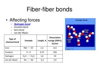
Connective tissue layers • Endomysium • Perimysium • Epimysium Purslow & Trotter 1994
Structural definition • Qualitative • Epimysium • Discrete tendon • Insertion (gastroc) • Origin (extensor digiti longus) • Easy to separate • Electrophysiological • Common nerve • Common reflex
3-D structures • Curved (centroid) paths • Curved fiber paths • Distributed attachments • Varying fascicle length
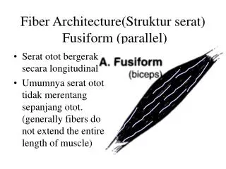
Categorizing • Pennation • Longitudinal • Unipennate • Bipennate • Multipennate • Approximation • Fascicle length • Force capacity
Historical • Stensen (1660) • Borelli (1680) • Gosch (1880)
Idealized muscles • Muscle mass (M) • Muscle length (Lm) • Fascicle length (Lf) • Pennation angle (q) • “Physiological” cross sectional area (PCSA)
The Gans & Bock Model • Vastus Intermedius • Identical facsicles • Originate directly from bone • Insert into tendon that lies parallel to bone • Geometrical constraints • Tendon moves parallel to bone • Constant volume • 2-D approximation • No change “into the paper” • Constant area
Force capacity d • Physiological cross-sectional area • Sum fascicles perpendicular to axis • Not measurable • Fm = Ts * PCSA • Prism approximation • Volume = b*d • B sin(q) = V/Lf • PCSA = V/Lf =M/r/Lf • Project force to tendon • Ft = Fm cos(q) = Ts*M/r/Lf * cos(q) b q Fm Lf PCSA Ft
Test PCSA • Spector & al., 1980 • Cat soleus and medial gastrocnemius • Powell & al., 1984 • Guinnea pig: 8 calf muscles 2.5 Powell Soleus 2.0 MG Spector 1.5 130% 1.0 41% Predicted PCSA (●) Predicted Ft (o) 0.5 6% 0.7% Relative measure 0.0 Po Po/g Po/pcsa Po/Ft Measured force
Are pennate muscles strong? • Ft = Ts*M/r/Lf * cos(q) • cos(q) is always ≤1 • Ft ≤ Fm • Fiber packing • Series sarcomeres (A=1, F=1) • Parallel sarcomeres (A=6, F=6) • Pennatesarcomeres (A = 6, F=5.2)
Length change d • Fiber shortens from ff1 • Rotates from q q1 • b*d constant • b*f*sin(q) = b*f1*sin(q1) • h = f*cos(q)-f1*cos(q1) • Fractional shortening in muscle isgreater than the fractional shorteningof fascicles • If the fascicles rotate much • eg: 15° fibers, fascicle shorten 25%muscle 27% f1 b h q q1 f
Operating range • Muscle can shorten ~50% (Weber, 1850) • Operating range proportional to length • Spasticity • Reduced mobility (Crawford, 1954) • Length-tension relationship • Useful range stronglydependent on Lo • Pennate fibers shortenless than their muscle
Velocity • Force-velocity relationship • Shortening muscle produces less force • Power = force * speed • Acceleration • Architecture andbiochemistry influenceVmax • Fiber type: 2x • Fiber length: 12x
Other Geometries • Point origin, point insertion • Elastic aponeurosis • Increase length with force • Vm = Va + Vf • Multipennate muscles Cos(a-q) Cos(a) Cos(q) Cos(a)
Other subdivisions • Multiple bellies • Digit flexors/extensors • Biceps/Triceps • Multiple discrete attachments • Compartments • Most “large” muscles • Internal connective tissue • Internal nerve branches
Multiple bellies • Rat EDL • 4 insertion tendons • 2 nerve branches • Glycogen depletion • Discrete branch territories • Mixing at ventral root Balice-Gordon & Thompson 1988
Compartments • Cat lateral gastrocnemius • Dense internal connective tissue • Surface texture • Internal nerve branches English & Ledbetter, 1982
LG Compartments • Motor unit • Axon+innervated fibers • Constrained tocompartment English & Weeks, 1984
Neural view • Does NS use the same divisions as anatomists? • Careful training can control single motoneuron • Behavioral recruitment spans muscles • Mechanical tuning • Training
Anatomical vs neural division • Muscle • Easily separated • Separately innervated • Multi-belly • Partly separable • Slight overlap of nerve territories • Compartment • Inseparable • Slight overlap of nerve territories
Fibers and fascicles • Rodents • Fiber = fascicle • Easiest experimental model • Small animals • Fascicle 5-10 cm • Fiber 1-2 cm (conduction velocity ~2-5 m/s)
Motor unit distribution Fibers innervated by single MN are near one MEP band • MU localized longitudinal Motor endplates in sternomanibularis Smits et al., 1994 Purslow & Trotter, 1994
3-D reconstruction • Relatively straight fibers • Taper-in, taper-out 1 mm Ounjian et al., 1991
Mechanical independence • Bag of spaghetti model • Independent muscle/belly/compartment/fiber • Little force sharing • Fiber composite model • Adjacent structures coupled elastically • Lateral force transmission
Fiber level force transmission • Sybil Street, 1983 • Frog sartorius • All but one fiber removed from half muscle • Anchor remaining fiber ends • Anchor segment and “clot” • Same force
“Belly” level force transmission • Huijing & al., 2002 • Rat EDL • Separate digit tendons • Cut one-by-one (TT) • Pull bellies apart (MT) • Little force change withtenotomy only
Muscle level force transmission • Maas & al., 2001 • Rat TA and EDL • Separate controlof muscle lengths • Measure both EDLorigin&insert F • 10% EDL-TA trans
Summary • Architectural quantification: M, Lm, Lf, q • Estimates of force production: PCSA (Fm), Ft • Simple models are “pretty good” • Sub-muscular structures: compartments • Neural structure is not the same as muscle structure