Download
1 / 90
1.08k likes | 2.44k Views
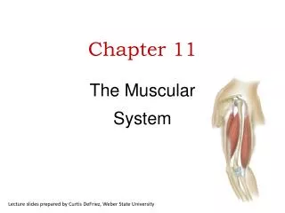
Chapter 11: The Muscular System. The Muscular System. Consists only of skeletal muscles Muscle organization affects power, range, and speed of muscle movement. Fascicles. Muscle cells ( fibers ) are organized in bundles ( fascicles ) Classification of Skeletal Muscles

E N D
The Muscular System • Consists only of skeletal muscles • Muscle organization affects power, range, and speed of muscle movement
Fascicles • Muscle cells (fibers) are organized in bundles (fascicles) • Classification of Skeletal Muscles • By the way fascicles are organized • By relationships of fascicles to tendons
Muscle Organization • Groups of fibers are organized into fascicles • Fibers in fascicle run parallel to fascicle, but fascicle can be arranged in 4 different shapes with respect to tendon: • Parallel Muscles • Convergent Muscles • Pennate Muscles - Unipennate, Bipennate, Multipennate • Circular Muscles
1. Parallel Muscles • Fascicles run parallel to length of the muscle • Most skeletal muscles are arranged this way • Able to change length extensively • Can move load over a great distance Figure 11–1a
Parallel Muscle Body • The center or body of the muscle thickens when parallel muscle contracts • Tension • Depends on total number of myofibrils • Directly relates to cross section of muscle • 1 in.2 (6.45 cm2) of cross section develops 50 lb (23 kg) of tension
2. Pennate Muscles • Fascicles are arranged at an angle to tendon A. Unipennate: Fascicle angled on one side of tendon B. Bipennate: Tendon in middle with angled fascicles on either side C. Multipennate: - Branched tendon with fascicles organized around each branch **Pennate muscles produce more tension than parallel muscles but cannot move so far, less distance produced • Unipennate B. Bipennate C. Multipennate
Pennate Muscles • Unipennate: • fibers on 1 side of tendon • e.g., extensor digitorum • Bipennate: • fibers on both sides of tendon • e.g., rectus femoris • Multipennate: • tendon branches within muscle • e.g., deltoid
3. Convergent Muscles • Fascicles spread out like a fan on one end and converge to a single point on the other • Produce less tension and distance than parallel muscles but • Independent contraction of fascicles can produce different movement from the same muscle • Provides versatility • Muscle fibers pull in different directions, depending on stimulation Figure 11–1b
4. Circular Muscles • Also called sphincters • Concentric arrangement of fascicles • Function: • Decrease diameter of openings to guard entrances and exits • e.g., obicularis oris Figure 11–1f
Why does a pennate muscle generate more tension than does a parallel muscle of the same size? Parallel fibers do not respond to calcium. A pennate muscle contains more muscle fibers. Muscle force is concentrated on the insertion in pennate muscles. This is not a true statement.
Which type of muscle would you expect to be guarding the opening between the stomach and the small intestine? convergent muscle multipennate muscle parallel muscle circular muscle (sphincter)
Muscle Terminology • Muscles have 1 fixed point of attachment (origin) and 1 moving point of attachment (insertion) • Origin: • Where the fixed end of the muscle attached to bone, cartilage, or CT • Origin is usually proximal to insertion • Insertion: • Where the moveable end attaches • Action: • The specific movement produced by the muscle during contraction • e.g., flexion, extension, adduction, etc.
Muscle Interactions • Muscles work in groups to maximize efficiency • Smaller muscles reach maximum tension first, followed by larger, primary muscles
Muscle Terminology • Muscle often work in groups to increase tension or fine tune movement • Different muscles serve different function: • Agonist: • prime mover, muscle most responsible for the movement • Synergist: • a muscle with the same action as the agonist • assists agonist at the beginning of contraction when fiber length is not optimal for agonist • helps start motion or stabilize origin of agonist (fixator) • Antagonist: • a muscle whose action opposes the agonist • produces the opposite action to fine tune movement by the agonist
Muscle Opposition • Agonists and antagonists work in pairs: • when 1 contracts, the other stretches • i.e. flexors–extensors abductors–adductors
The name of a muscle helps identify its location, appearance, or function.
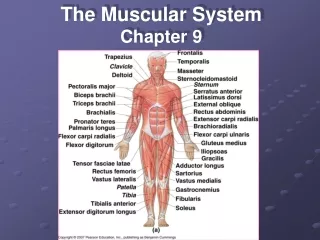
Descriptive Names for Skeletal Muscles • Location in the body • Fascicle organization • Relative position • Structure, Size and Shape • Origin and insertion • Action
Naming Muscles • Names of muscle are derived from aspects of their features: • Location: - Named for part of the body where they’re located - e.g. Brachii, Abdominis • Fascicle Organization: - Named for how fascicles are organized with respect to the body - e.g. Rectus = straight Oblique = angle • Relative Position: - Named for depth when layered - e.g. Externus/Superficialis = top, surface Internus/Profundus = deep
Naming Muscles 4. Structure, Size and Shape: • Number of tendons - e.g. triceps, biceps • Shape of Muscle • e.g. trapezius = trapezoid deltoid = triangle soleus = fish • Size of muscle relative to others • Major = bigger • Maximus = biggest • Longus = long • Vastus = great
Naming Muscles • Origin and Insertion: - Name of regions of attachment, origin first, insertion second - e.g. sternocleidomastoid Origin = manubrium of sternum and medial clavicle Insertion = mastoid process 6. Action: - Named for action performed and region acted upon - e.g. extensor digitorum Usually multiple naming schemes are combined to name the muscle: e.g. flexor carpi ulnaris * Individual muscles, orgins, insertions and actions are examined in lab.
Naming Skeletal Muscles Table 11–1 (1 of 2)
Naming Skeletal Muscles Table 11–1 (2 of 2)
Effects of Aging on the Muscular System • Skeletal muscle fibers become smaller in diameter • Skeletal muscles become less elastic: • develop increasing amounts of fibrous tissue (fibrosis) • Decreased tolerance for exercise • Decreased ability to recover from muscular injuries
Muscle A abducts the humerus, and muscle B adducts the humerus. What is the relationship between these two muscles? synergists antagonists agonists fixators
What does the name flexor carpi radialis longus tell you about this muscle? its size its function its location 1, 2, and 3 are correct
A Closer Look at the Muscular System
Axial and Appendicular Muscles Figure 11–3a
Axial and Appendicular Muscles Figure 11–3b
Divisions of the Muscular System • Axial muscles: • position head and spinal column • move rib cage • 60% of skeletal muscles • Appendicular muscles: • support pectoral and pelvic girdles • support limbs • 40% of skeletal muscles
The Axial Muscles • Divisions based on location and function: • muscles of head and neck • muscles of vertebral column • oblique and rectus muscles • muscles of pelvic floor
Muscles of Facial Expression Figure 11–4b
Summary: Muscles of Facial Expression Table 11–2 (1 of 2)
Summary: Muscles of Facial Expression Table 11–2 (2 of 2)
Anterior Muscles of the Neck Figure 11–9
Oblique and Rectus Muscles • Lie within the body wall Figure 11–11a, b
Functions of Oblique and Rectus Muscles • Oblique muscles: • compress underlying structures • rotate vertebral column • Rectus muscles: • flex vertebral column
Oblique Muscles • Thoracic region: • intercostal muscles (external and internal intercostals): • respiratory movements of ribs • Abdominopelvic region (same pattern as thoracic): • external oblique muscles • internal oblique muscles
Rectus Group • Rectus abdominis: • between xiphoid process and pubic symphysis • divided transversely by tendinous inscriptions
Oblique Muscles Table 11–9 (1 of 2)
Oblique and Rectus Muscles Table 11–9 (2 of 2)
The structures and functions of the major muscle groups of the upper and lower limbs.
The Appendicular Muscles Figure 11–13b
The Appendicular Muscles Figure 11–13a
The Appendicular Muscles • Position and stabilize pectoral and pelvic girdles • Move upper and lower limbs • Move the arm • Move the forearm and hand • Move the hand and fingers
Muscles that Position the Pectoral Girdle Figure 11–14b
Muscles that Position the Pectoral Girdle Figure 11–14a
Muscles that Position the Pectoral Girdle • Trapezius: • superficial • covers back and neck to base of skull • inserts on clavicles and scapular spines • Rhomboid and levator scapulae: • deep to trapezius • attach to cervical and thoracic vertebrae • insert on scapular border • Serratus anterior: • on the chest • originates along ribs • inserts on anterior scapular margin
Muscles that Position the Pectoral Girdle Tables 11–11