Download
1 / 22
240 likes | 512 Views
Vascular Access How I Do It. Gareth Griffiths Department of Vascular Surgery, Ninewells Hospital, Dundee, UK Chairman of the Specialty Advisory Committee in General Surgery. Team Working. Nephrologist Vascular technician Dialysis nurses Radiologist Anaesthetist Surgeon.

E N D
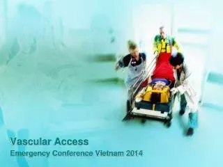
Vascular Access How I Do It Gareth Griffiths Department of Vascular Surgery, Ninewells Hospital, Dundee, UK Chairman of the Specialty Advisory Committee in General Surgery
Team Working • Nephrologist • Vascular technician • Dialysis nurses • Radiologist • Anaesthetist • Surgeon
Pre Operative Assessment • Clinical • Ultrasound • Vein • Size • Intravenous webs / thrombus • Beware of spasm • Examine full length • Central deep veins • Artery • Wall calcification • Waveform pattern • Examine full length
Pre Operative Assessment • Suspicion of central vein stenosis • Venography • MR • CT • Catheter
Pre Operative Assessment • Upper limb before lower limb • Non-dominant before dominant • Distal before proximal • Autogenous before prosthetic • Priority depends on the patient • Already on haemodialysis • Age
Sequence of Operations • Radio-cephalic • Brachio-cephalic • Brachio-basilic • Brachio-axillary PTFE • Long saphenous thigh straight / loop • PTFE thigh straight / loop • Superficial femoral vein straight / loop
Rarely Needed Options • Iliac artery – vein PTFE loop • Necklace axillary artery – vein PTFE • Arterio-arterial PTFE
Operative Technique • Local or regional anaesthesia • Gentle handling of tissues • Meticulous technique • Microvascular instruments • Magnification and light
Operative Technique • Radiocephalic / brachiocephalic • Single incision when possible • Mobilise vein • Avoid twisting • Isolate artery • Microvascular clamps • Careful siting of arteriotomy • Relieve spasm – hydrostatic pressure, balloon • Check, check, check
Operative Technique • Brachiobasilic • One stage procedure • Mobilise maximum length of vein • 3-4 short incisions ?endoscopic • Fashion tunnel to match vein length • Straight tunnel preferable • Arched tunnel if vein short
Operative Technique • Prosthetic fistula • 6mm PTFE • Miller cuff at each end • Protects native vessels from PTFE thrombosis • Facilitates removal of infected PTFE
Follow Up • Life long surveillance • Clinical • Bleeding • Cannulation difficulties • Ultrasound • Duplex identified stenosis • Dialysis parameters • Venous pressure < 180mm Hg • Arterial pressure > -180 mmHg • Urea reduction ratio >70% • Access flow >600ml/min <25% fall
Multidisciplinary Meeting • Surgeon • Vascular technologist • Nephrologist • Radiologist • Dialysis specialist nurse • Discuss all patients with duplex or dialysis identified issues • Review all post intervention outcomes
Multidisciplinary Meeting • Selective intervention • Dialysis parameter abnormality + identified stenosis • Angioplasty first • Cutting balloon if necessary • Stenting if necessary • Surgical re-fashioning • Failed endovascular intervention
Multidisciplinary Meeting • Surveillance and repeated intervention • Longest assisted primary patency possible • Pre-emptive new fistula creation • When fistula failure is predicted • Before loosing fistula access
Fistula Thrombosis • Attempt salvage unless • Fistula had been identified as failing • Active infection • Aneurysmal with organised thrombus • Radiological salvage • Combined mechanical and lytic • Concomitant angioplasty / stenting when needed • Surgical thrombectomy • Early post op thrombosis
Aneurysmal Fistulae • No issue if uncomplicated • Cosmetic • Ask patient to accept • Thin skin • Lateral cannulation • Bleeding • Repair with vein buttress occasionally possible • Ligation often needed
Steal • Exclude central arterial stenosis • Often mild • Conservative management • Significant • Pain, tissue loss • High flow • Good distal vessels • High fistula flow • Low flow • Diseased distal vessels • Critical flow distally
Steal • High flow • Assess direction of flow in distal artery • If retrograde • Radial fistula – ligate distal radial artery • Maximises ulnar flow into hand • Brachial fistula - Distal Revascularisation and Interval Ligation (DRIL) • Restores antegrade flow towards hand • If antegrade • Band fistula • Reduces fistula flow, improves distal perfusion
Steal • Low flow • Diseased distal vessels • Poor outlook • Increasingly common • Banding • Rarely possible – fistula flow already low • Ligate fistula
Swollen Arm • Assess promptly • Generally indicates central vein stenosis • Urgent catheter venography • Angioplasty or stenting when possible • May need fistula ligation • Try to avoid with careful pre-op assessment
Vascular AccessHow I Do It • Multi-disciplinary team • Attention to detail • Logical sequence for fistula creation • Perseverance • Fistula surveillance and repeated interventions