Download
1 / 53
540 likes | 770 Views
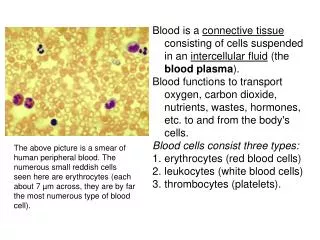
DISEASES OF WHITE BLOOD CELLS Premed 3 Dr Roopa. What Is Leukemia?. Cancer of the white blood cells Acute or Chronic Affects ability to produce normal blood cells Bone marrow makes abnormally large number of immature white blood cells called blasts.

E N D
DISEASES OF WHITE BLOOD CELLS Premed 3 Dr Roopa
What Is Leukemia? • Cancer of the white blood cells • Acute or Chronic • Affects ability to produce normal blood cells • Bone marrow makes abnormally large number of immature white blood cells called blasts
History • Means “white blood” in Greek • Discovered by Dr. Alfred Velpeau in France, 1827 • Named by pathologist Rudolf Virchow in Germany, 1845
↑ leukocytes Acute leukemias 1. Acute Lymphoblastic Leukemia (ALL) 2. Acute Myelogenous Leukemia (AML) Chronic leukemias 1.Chronic Lymphoblastic Leukemia (CLL) 2. Chronic Myelogenous Leukemia (CML) Leukemias
Myeloid tissue is a biologic tissue with the ability to perform hematopoiesis. It is mainly found as the red bone marrow in bones, and is often synonymous with this. However, myeloid can also be present in the liver and spleen . • A myelocyte is a young cell of the granulocytic series, occurring normally in bone marrow, but not in circulating blood (except when caused by certain diseases).
Myelogenous Leukemia • Leukemia characterized by proliferation of myeloid tissue (as of the bone marrow and spleen) and an abnormal increase in the number of granulocytes, myelocytes, and myeloblasts in the circulating blood
Granulocytes are a category of white blood cells characterized by the presence of granules in their cytoplasm They are also called polymorphonuclear leukocytes (PMN or PML) because of the varying shapes of the nucleus, which is usually lobed into three segments. • The myeloblast is a unipotent stem cell, which will differentiate into one of the actors of the granular series.
Acute Leukemias • Blast predominate • Child or elder • Short & drastic course • ALL – Lymphoblasts (pre-B or pre-T) • AML – Myeloblasts
Chronic Leukemias • More mature cells • Middle age • Longer & less devastating course • CLL – Lymphocytes • CML – Myeloid stem cells
Acute Leukemias • accumulation of blasts in the marrow
Acute Lymphoblastic Leukemia (ALL) • Children • Lymphoblasts (pre-B or pre-T) • Neoplastic transformation of the lymphoid stem cells • Progressive accumulation of Lymphoblasts in the bone marrow • Suppression of normal hemopoiesis
• Primarily a disease of children and young adults • B-cell subtype (80%) • T-cell subtype (20%)
ALL - Pathogenesis • Etiology unknown • Genetic predisposition (some) Down syndrome • Translocations (worse prognosis) t(4;11)t(12;21)t(9;22)
Signs &Symptoms • Anemia • Infection • Bleeding • Bone pain • Arthritis • Splenomegaly • Lymphadenopathy • CNS involvement
ALL Prognosis: • Age 3-7, pre-B, L1 : > 90% - CR( 2/3 - cure ) • Adults, mature B and T-ALL: Less favorable Therapy: Chemotherapy ( w/CNS prophylaxis ), supportive care, BMT
ACUTEMYELOGENOUS LEUKEMIA ( AML ) • Adults • Myeloblasts • monoblasts, eosinoblasts, megakarioblasts, proerythroblasts, basophiloblasts • Auer rods in the cytoplasm of the cells • Very rapidly progressive malignancy
AML - Pathogenesis Environmental factors: High-dose radiation exposure Myelotoxic agents (benzene, alkylating agents) Genetic abnormalities: Down syndrome, Immunodeficiency diseases
Differentiation from ALL may be made by microscopy – presence of Auer Rods. Clinical features based on • Marrow failure –anemia, bleeding, DIC, infection… • Leukemic infiltration – bone pain, CNS signs, hepatosplenomegaly, lymphadenopathy… • Constitutional upset -- malaise, fever, weakness, polyarthritis.
Course: • Rapidly fatal if untreated (< 2 mo ) • Median survival - 3 years after chem.. • Adverse prognostic factors: Age > 60 t(9;21), Previous chemotherapy Leukocytosis > 100,000 /ul Therapy:Chemotherapy, supportive, BMT
***Remember this*** For Acute leukemias • acute leukemias = too many blasts in the marrow • 2 broad categories: AML vs. ALL • a hematologic urgency • prognosis is poor in adults; but good in kids with ALL.
CHRONIC LYMPHOCYTIC LEUKEMIA (CLL) A monoclonal lymphoproliferative disorder characterized by lymphocytosis(>4000/cu.mm), lymphadenopathy and splenomegaly B - CLL > 95% T - CLL
CLL •Most common adult leukemia in Western society (30% of all leukemias) • Monoclonal proliferation of the small lymphocytes… • Age > 40 M:F / 2:1
CLL PB and BM
CLL - Pathology Blood: • Lymphocytosis ( > 10,000 u/L - diagnostic ) • (+) Coombs test (20%) • Hypogammaglobulinemia (50-70%) • Anemia, thrombocytopenia, neutropenia Bone marrow: • nodular / interstitial infiltrates • diffuse - obliteration of normal hemopoiesis Lymphadenopathy,Hepatosplenomegaly(50-60%)
CLL - Clinical course Initially: asymptomatic Advanced disease: • bacterial infections, hemorrhage Prognostic factors: • extent of tumor burden • pattern of marrow infiltration • chromosomal abnormalities Median survival: ~ 6 years
CLL • One more peripheral blood findings in CLL is Presence of Smudge cells ( parachute cells). • Along with increased number of normal appearing lymphocytes.
Chronic Myelogenous Leukemia (CML) • Excessive development of mature neoplastic granulocytes in the bone marrow • Move into the peripheral blood in massive numbers • Ultimately infiltrate the liver and spleen
Chronic Myelogenous Leukemia • Philadelphia chromosome • 9 and 22 translocation almost specific to CML • Produces BCR/c-abl fusion oncogene • The chromosome abnormality that causes chronicmyeloidleukemia (CML) (9 &22) • Genetic marker • Chronic, stable phase followed by acute, aggressive (blastic) phase
FISH showing the BCR (green), ABL (orange), and BCR-ABL fusion signals (arrow): A=positive (contains a residual ABL signal), B=normal
i.e – mainly uncontrolled proliferation of myeloid cells. • Males more than females • Splenomegaly – sometimes massive..
**Philadelphia chromosome** • Hybrid chromosome with translocation between the long arm of chr. 9 and long arm of chr.22 . --- t(9:22). • May be present in granulocyte, RBC or platelet precursors in more than 95% of CML..
CML - Pathology Bone marrow: • Hypercellular / predominant granulocytic hyperplasia • Increased megakaryocytes (small forms) • Normal to decreased erythroid precursors Peripheral blood: • Granulocytosis (>25,000/cmm), with immature cells, • Basophilia,eosinophilia, Extramedullaryhemopoiesis: Spleen, liver, lymph nodes
CML - Clinical Features 15 - 20% of all leukemias; age 25-60 Symptoms: - non-specific - related to hypermetabolism (high cell turnover) - related to splenomegaly Course: - chronic phase (mean survival, 3-4y) - accelerated phase - blast crisis / myeloid or lymphoid (survival, < 1y) Therapy: chemotherapy; BMT
Hodgkin’s lymphoma • a lymphoid neoplastic disorder of B cell origin • A disease marked by chronic enlargement of the lymph nodes, often local at the onset and later generalized. • characterized by the presence of Reed-Sternberg cells (or variants of RS cells) in the affected tissues • Enlargement of the spleen and often of the liver is also present.
Reed-Sternberg cells • Considered to be a malignant neoplasm of lymphoid cells of uncertain origin. • Owl – Eye appearance • These cells have mirror-image nuclei. • Young adults and elderly
Signs & Symptoms • Enlarged painless nodes – mostly neck & axilla • Fever, wt.loss, pruritis & night sweats. • Pel – Ebstein Fever – fever alternating with long periods (15-28days). • Anemia • Cachexia • Hepatosplenomegaly
Hodgkin’s disease: Summary • a lymphoid neoplasm of B cell origin • characterized histologically by Reed Sternberg cells (or variants) • commonly presents with lymphadenopathy or mediastinal mass in young adults • treatment modality depends on stage • curable in most
Non Hodgkin's Lymphoma • These include all lymphomas without Reed-Sternberg cells. • Most are B-cell proliferations • Extra nodal involvement is there..
Signs & Symptoms • Often symptomless • Lymphadenopathy, wt.loss.. • Extra nodal spread – skin, bone, gut, CNS, lung.. • Pancytopenia may be there • Infections are very common. • Fatigue, unexplained fever, sweats • Enlarged Tonsils and adenoids
FOLLICULAR LYMPHOMA • Follicular lymphoma’s – most common in US • Derived from B lymphocytes • t (14:18) • Affects middle age • Not very aggressive – mean survival 7-10 yrs • Rarely they may transform in to aggressive type of lymphomas
Burkitt’s Lymphoma • A Lymphoblastic lymphoma mainly in African children. • Mostly associated with EBV infection. • Involves facial bones ( Jaw), ovaries, and abdominal lymph nodes. • Undifferentiated stem cells with scattered pale macrophages containing nuclear debris. • Isolated histiocytes on background of abnormal lymphoblasts (STARRY SKY APPERANCE) • t (8:14) • High incidence in AIDS pt’s