Download
1 / 15
150 likes | 418 Views
Review of Microscopes. Plant and animal cell structure. Types of microscopes. 3 types of microscopes Light Electron Scanning/tunnelling. Light microscopes. Microscope Care. Always carry with 2 hands Only use lens paper for cleaning Do not force knobs Always store covered

E N D
Review of Microscopes Plant and animal cell structure
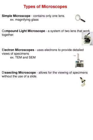
Types of microscopes • 3 types of microscopes • Light • Electron • Scanning/tunnelling
Microscope Care • Always carry with 2 hands • Only use lens paper for cleaning • Do not force knobs • Always store covered • Keep objects clear of desk and cords
Microscope parts Eyepiece BodyTube RevolvingNosepiece Arm ObjectiveLens Stage StageClips CoarseFocus Diaphragm FineFocus Light Base
Using the microscope • Place the Slide on the Microscope • Use Stage Clips • Click Nosepiece to the lowest (shortest) setting • Look into the Eyepiece • Use the Coarse Focus
Using high power • Follow steps to focus using low power • Click the nosepiece to the longest objective • Do NOTuse the Coarse Focusing Knob • Use the Fine Focus Knob to bring the slide What can you find on your slide?
Task 1 • Set up the microscope to view the prepared slides • View at least 4 different slides, make a labelled diagram of what you see • Classify your cells as being either plant or animal cells • Write a simple experimental write up for this experiment
Write up • Needs the following heading Aim: The purpose of the experiment Materials: A list of what was used Procedure: A step by step description of the process Results: Your labelled diagrams and any other observations Discussion: Analysis of results, include similarities and differences between animal and plant cells from your observations, include any errors of difficulties. Conclusion: Write a concluding paragraph about the results and the aim
Electron Microscopy (TEM) • beams of electrons are used to produce images • wavelength of electron beam is much shorter than light, resulting in much higher resolution with this type of microscope
The Scanning Electron Microscope (SEM) • uses electrons reflected from the surface of a specimen to create an image • produces a 3-dimensional image of specimen’s surface features