Download
1 / 5
50 likes | 111 Views
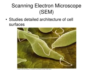
Explore normal vs. osteoporotic bone architecture in lumbar vertebrae using low-power SEM images. Witness bone erosion and osteoclast activity in 30-year-old and 71-year-old subjects.

E N D
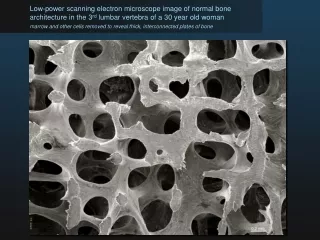
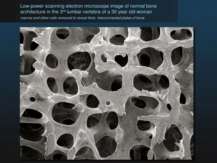
Low-power scanning electron microscope image of normal bone architecture in the 3rd lumbar vertebra of a 30 year old woman marrow and other cells removed to reveal thick, interconnected plates of bone
0.2 mm Low-power scanning electron micrograph of osteoporotic bone architecture in the 3rd lumbar vertebra of a 71 yr old woman marrow and other cells removed to reveal eroded bone elements
Low-power scanning electron micrograph of osteoporotic bone architecture in the 3rd lumbar vertebra of a 71 yr old woman marrow and other cells removed to reveal eroded bone elements
Scanning electron micrograph of a rat osteoclast and resorption pit on a bone surface