Download
1 / 57
570 likes | 682 Views
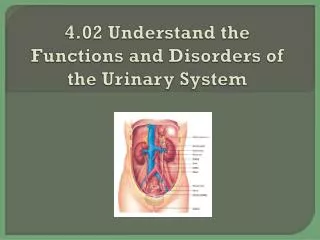
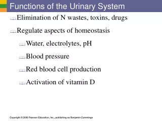
Functions of the Urinary System. Elimination of waste products Nitrogenous wastes Toxins Drugs. Functions of the Urinary System. Regulate aspects of homeostasis Water balance Electrolytes Acid-base balance in the blood Blood pressure Red blood cell production Activation of vitamin D.

E N D
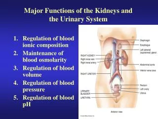
Functions of the Urinary System • Elimination of waste products • Nitrogenous wastes • Toxins • Drugs
Functions of the Urinary System • Regulate aspects of homeostasis • Water balance • Electrolytes • Acid-base balance in the blood • Blood pressure • Red blood cell production • Activation of vitamin D
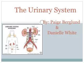
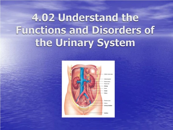
Organs of the Urinary System • Kidneys • Ureters • Urinary bladder • Urethra
Location of the Kidneys • Against the dorsal body wall in a retroperitoneal position (behind the parietal peritoneum) • At the level of the T12 to L3 vertebrae • The right kidney is slightly lower than the left (due to position of the liver)
12th rib (b) Figure 15.1b
Hepatic veins (cut) Inferior vena cava Renal artery Adrenal gland Renal hilum Aorta Renal vein Kidney Iliac crest Ureter Rectum (cut) Uterus (part of female reproductive system) Urinary bladder Urethra (a) Figure 15.1a
Regions of the Kidney • Renal cortex—outer region • Renal medulla—inside the cortex • Renal pelvis—inner collecting tube
Kidney Structures • Renal or medullary pyramids—triangular regions of tissue in the medulla • Renal columns—extensions of cortex-like material inward that separate the pyramids • Calyces—cup-shaped structures that funnel urine towards the renal pelvis
Renal column Major calyx Renal cortex Minor calyx Renal pyramid (a) Figure 15.2a
Cortical radiate vein Cortical radiate artery Arcuate vein Arcuate artery Renal column Interlobar vein Interlobar artery Segmental arteries Renal cortex Renal vein Renal artery Minor calyx Renal pelvis Major calyx Renal pyramid Ureter Fibrous capsule (b) Figure 15.2b
Blood Supply • One-quarter of the total blood supply of the body passes through the kidneys each minute • Renal artery provides each kidney with arterial blood supply
Nephron Anatomy and Physiology • The structural and functional units of the kidneys • Responsible for forming urine • Main structures of the nephrons • Glomerulus • Renal tubule
Nephron Anatomy • Glomerulus • Knot of capillaries • Glomerulus sits within a glomerular (Bowman’s) capsule (the first part of the renal tubule)
Nephron Anatomy • Renal tubule extends from glomerular capsule and ends at the collecting duct • Glomerular (Bowman’s) capsule • Proximal convoluted tubule (PCT) • Loop of Henle • Distal convoluted tubule (DCT)
Nephron Anatomy • Nephrons are associated with two capillary beds • Glomerulus • Peritubular capillary bed
Cortical nephron Fibrous capsule Renal cortex Collecting duct Renal medulla Renal cortex Proximal convoluted tubule Renal pelvis Glomerulus Ureter Juxtamedullary nephron Distal convoluted tubule Loop of Henle Renal medulla (a) Figure 15.3a
Proximal convoluted tubule (PCT) Peritubular capillaries Glomerular capillaries Distal convoluted tubule (DCT) Glomerular (Bowman’s) capsule Efferent arteriole Afferent arteriole Cells of the juxtaglomerular apparatus Cortical radiate artery Arcuate artery Arcuate vein Cortical radiate vein Collecting duct Loop of Henle (b) Figure 15.3b
Collecting Duct • Receives urine from many nephrons • Run through the medullary pyramids • Deliver urine into the calyces and renal pelvis
Proximal convoluted tubule (PCT) Peritubular capillaries Glomerular capillaries Distal convoluted tubule (DCT) Glomerular (Bowman’s) capsule Efferent arteriole Afferent arteriole Cells of the juxtaglomerular apparatus Cortical radiate artery Arcuate artery Arcuate vein Cortical radiate vein Collecting duct Loop of Henle (b) Figure 15.3b
Three steps to Urine Formation • Glomerular filtration • Tubular reabsorption • Tubular secretion
Glomerulus physiology • Fed and drained by arterioles • Afferent arteriole—arises from a cortical radiate artery and feeds the glomerulus • Efferent arteriole—receives blood that has passed through the glomerulus • Specialized for filtration • High pressure forces fluid and solutes out of blood and into the glomerular capsule
Glomerular capsular space PCT Glomerular capillary covered by podocytes Efferent arteriole Afferent arteriole (c) Figure 15.3c
Glomerular Filtration • Nonselective passive process • Water and solutes smaller than proteins are forced through capillary walls • Proteins and blood cells are normally too large to pass through the filtration membrane • Filtrate is collected in the glomerular capsule and leaves via the renal tubule
Tubular Reabsorption • The peritubular capillaries reabsorb useful substances • Water • Glucose • Amino acids • Ions • Some reabsorption is passive, most is active • Most reabsorption occurs in the proximal convoluted tubule
Tubular Reabsorption • What materials are not reabsorbed? • Nitrogenous waste products • Urea—protein breakdown • Uric acid—nucleic acid breakdown • Creatinine—associated with creatine metabolism in muscles
Tubular Secretion: Reabsorption in Reverse • Some materials move from the blood of the peritubular capillaries into the renal tubules • Hydrogen and potassium ions • Creatinine • Process is important for getting rid of substances not already in the filtrate • Materials left in the renal tubule move toward the ureter
Afferent arteriole Glomerular capillaries Efferent arteriole Cortical radiate artery Glomerular capsule 1 Rest of renal tubule containing filtrate Peritubular capillary 2 3 To cortical radiate vein Three major renal processes: Urine Glomerular filtration: Water and solutes smaller than proteins are forced through the capillary walls and pores of the glomerular capsule into the renal tubule. 1 Tubular reabsorption: Water, glucose, amino acids, and needed ions are transported out of the filtrate into the tubule cells and then enter the capillary blood. 2 Tubular secretion: H+, K+, creatinine, and drugs are removed from the peritubular blood and secreted by the tubule cells into the filtrate. 3 Figure 15.4
Proximal convoluted tubule (PCT) Peritubular capillaries Glomerular capillaries Distal convoluted tubule (DCT) Glomerular (Bowman’s) capsule Efferent arteriole Afferent arteriole Cells of the juxtaglomerular apparatus Cortical radiate artery Arcuate artery Arcuate vein Cortical radiate vein Collecting duct Loop of Henle (b) Figure 15.3b
Characteristics of Urine • In 24 hours, about 1.0 to 1.8 liters of urine are produced • Urine and filtrate are different • Filtrate contains everything that blood plasma does (except proteins) • Urine is what remains after the filtrate has lost most of its water, nutrients, and necessary ions through reabsorption • Urine contains nitrogenous wastes and substances that are not needed
Characteristics of Urine • Yellow color due to the pigment urochrome (from the destruction of hemoglobin) and solutes • Dilute urine is a pale, straw color • Sterile • Normal pH of around 6 • Specific gravity of 1.001 to 1.035
Characteristics of Urine • Solutes normally found in urine • Sodium and potassium ions • Urea, uric acid, creatinine • Ammonia • Bicarbonate ions
Characteristics of Urine • Solutes NOT normally found in urine • Glucose • Blood proteins • Red blood cells • Hemoglobin • White blood cells (pus) • Bile
Ureters • Slender tubes attaching the kidney to the bladder • Continuous with the renal pelvis • Enter the posterior aspect of the bladder
Hepatic veins (cut) Inferior vena cava Renal artery Adrenal gland Renal hilum Aorta Renal vein Kidney Iliac crest Ureter Rectum (cut) Uterus (part of female reproductive system) Urinary bladder Urethra (a) Figure 15.1a
Urinary Bladder • Smooth, collapsible, muscular sac • Temporarily stores urine • Trigone—triangular region of the bladder base • Three openings • Two from the ureters • One to the urethra • In males, the prostate gland surrounds the neck of the bladder
Urinary bladder Ureter Internal urethral orifice Ureteral orifice Trigone External urethral sphincter Internal urethral sphincter Urogenital diaphragm Urethra Figure 15.6
Urinary Bladder Wall • Three layers of smooth muscle collectively called the detrusor muscle • Mucosa made of transitional epithelium • Walls are thick and folded in an empty bladder • Bladder can expand significantly without increasing internal pressure
Urinary Bladder Capacity • A moderately full bladder is about 5 inches long and holds about 500 mL of urine • Capable of holding twice that amount of urine
Umbilicus Superior wall of distended bladder Superior wall of empty bladder Pubic symphysis Figure 15.7
Urethra • Thin-walled tube that carries urine from the bladder to the outside of the body by peristalsis • Release of urine is controlled by two sphincters • Internal urethral sphincter • Involuntary and made of smooth muscle • External urethral sphincter • Voluntary and made of skeletal muscle
Urinary bladder Ureter Internal urethral orifice Ureteral orifice Trigone External urethral sphincter Internal urethral sphincter Urogenital diaphragm Urethra Figure 15.6
Urethra Gender Differences • Length • Females is 3 to 4 cm (1 inch) • Males is 20 cm (8 inches) • Location • Females—anterior to the vaginal opening • Males—travels through the prostate and penis
Urethra Gender Differences • Function • Females—only carries urine • Males—carries urine and is a passageway for sperm cells and semen
Micturition (Voiding) • Both sphincter muscles must open to allow voiding • The internal urethral sphincter is relaxed after stretching of the bladder • Pelvic splanchnic nerves initiate bladder to go into reflex contractions • Urine is forced past the internal urethra sphincter and the person feels the urge to void • The external urethral sphincter must be voluntarily relaxed to void
Fluid, Electrolyte, and Acid-Base Balance • Kidneys have four roles in maintaining blood composition • Excretion of nitrogen-containing wastes (previously discussed) • Maintaining water balance of the blood • Maintaining electrolyte balance of the blood • Ensuring proper blood pH
Maintaining Water Balance • Normal amount of water in the human body • Young adult females = 50 percent • Young adult males = 60 percent • Babies = 75 percent • The elderly = 45 percent • Water is necessary for many body functions, and levels must be maintained
Distribution of Body Fluid • Intracellular fluid (ICF) • Fluid inside cells • About two-thirds of body fluid • Extracellular fluid (ECF) • Fluids outside cells that includes • Interstitial fluid • Blood plasma
The Link Between Water and Salt • Solutes in the body include electrolytes like sodium, potassium, and calcium ions • Changes in electrolyte balance causes water to move from one compartment to another • Alters blood volume and blood pressure • Can impair the activity of cells
Maintaining Water Balance • Water intake must equal water output • Sources for water intake • Ingested foods and fluids • Water produced from metabolic processes • Thirst mechanism is the driving force for water intake