Download
1 / 17
170 likes | 468 Views
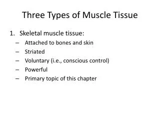
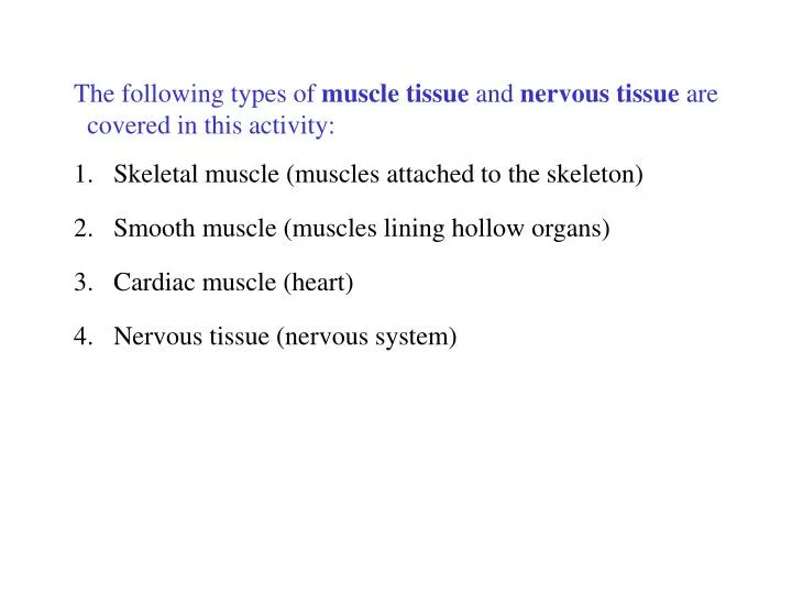
The following types of muscle tissue and nervous tissue are covered in this activity: 1. Skeletal muscle (muscles attached to the skeleton) 2. Smooth muscle (muscles lining hollow organs) 3. Cardiac muscle (heart) Nervous tissue (nervous system).

E N D
The following types of muscle tissue and nervous tissue are • covered in this activity: • 1. Skeletal muscle (muscles attached to the skeleton) • 2. Smooth muscle (muscles lining hollow organs) • 3. Cardiac muscle (heart) • Nervous tissue (nervous system)
Various membranes are also covered: • Cutaneous membranes • Mucous membranes • Serous membranes (parietal and visceral layers) • a. Peritoneum – abdominal organs • b. Pericardium – heart • c. Pleura – lungs
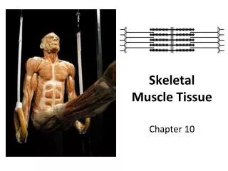
Figure 4.10a Muscle tissues. (a) Skeletal muscle Description: Long, cylindrical, multinucleate cells; obvious striations. Striations Function: Voluntary movement; locomotion; manipulation of the environment; facial expression; voluntary control. Nuclei Location: In skeletal muscles attached to bones or occasionally to skin. Part of muscle fiber (cell) Photomicrograph: Skeletal muscle (approx. 460x). Notice the obvious banding pattern and the fact that these large cells are multinucleate.
Figure 4.10b Muscle tissues. (b) Cardiac muscle Description: Branching, striated, generally uninucleate cells that interdigitate at specialized junctions (intercalated discs). Striations Intercalated discs Function: As it contracts, it propels blood into the circulation; involuntary control. Location: The walls of the heart. Nucleus Photomicrograph: Cardiac muscle (500X); notice the striations, branching of cells, and the intercalated discs.
Figure 4.10c Muscle tissues. (c) Smooth muscle Description: Spindle-shaped cells with central nuclei; no striations; cells arranged closely to form sheets. Smooth muscle cell Function: Propels substances or objects (foodstuffs, urine, a baby) along internal passage- ways; involuntary control. Location: Mostly in the walls of hollow organs. Nuclei Photomicrograph: Sheet of smooth muscle (200x).
Figure 4.9 Nervous tissue. Nervous tissue Description: Neurons are branching cells; cell processes that may be quite long extend from the nucleus-containing cell body; also contributing to nervous tissue are nonirritable supporting cells (not illustrated). Nuclei of supporting cells Cell body Neuron processes Axon Dendrites Cell body of a neuron Function: Transmit electrical signals from sensory receptors and to effectors (muscles and glands) which control their activity. Neuron processes Location: Brain, spinal cord, and nerves. Photomicrograph: Neurons (350x)
Figure 4.11 Classes of membranes. Mucosa of nasal cavity Cutaneous membrane (skin Mucosa of mouth Esophagus lining Mucosa of lung bronchi (a) Cutaneous membrane (the skin) covers the body surface. (b) Mucous membranes line body cavities open to the exterior. Parietal pleura Parietal peritoneum Visceral pleura Visceral peritoneum Visceral pericardium Parietal pericardium (c) Serous membranes line body cavities closed to the exterior.
Figure 4.11a Classes of membranes. Cutaneous membrane (skin) (a) Cutaneous membrane (the skin)covers the body surface
Figure 4.11b Classes of membranes. Mucosa of nasal cavity Mucosa of mouth Esophagus lining Mucosa of lung bronchi (b) Mucous membranes line body cavitiesopen to the exterior
Figure 4.11c Classes of membranes. Parietal peritoneum Parietal pleura Visceral pleura Visceral peritoneum Parietal pericardium Visceral pericardium (c) Serous membranes line body cavitiesclosed to the exterior
What kind of tissue does this represent? Skeletal muscle Where in the body can you find this tissue? muscles attached to the skeleton
What kind of tissue does this represent? Smooth muscle Where in the body can you find this tissue? muscles lining hollow organs
What kind of tissue does this represent? Cardiac muscle Where in the body can you find this tissue? heart
What kind of tissue does this represent? Nervous tissue Where in the body can you find this tissue? nervous system
Figure 4.11a Classes of membranes. Cutaneous membrane (skin) Membrane covers thebody surface
Figure 4.11b Classes of membranes. Mucosa of nasal cavity Mucosa of mouth Esophagus lining Mucosa of lung bronchi Membranes line body cavitiesopen to the exterior
Figure 4.11c Classes of membranes. Parietal pleura Parietal peritoneum Visceral pleura Visceral peritoneum Parietal pericardium Visceral pericardium Membranes line body cavitiesclosed to the exterior