Download
1 / 19
250 likes | 899 Views
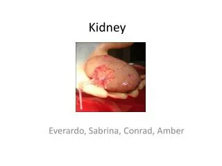
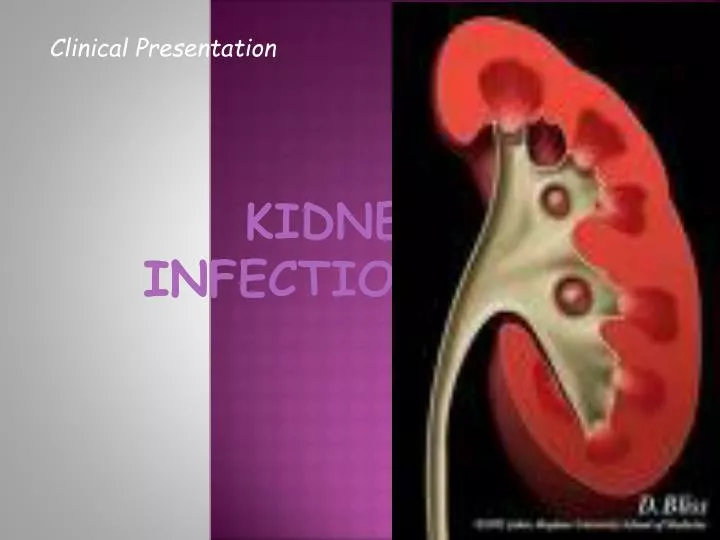
Clinical Presentation. KIDNEY INFECTION. Acute Pyelonephritis. Inflammation Kidney Renal pelvis. Acute Pyelonephritis Presentation & Findings. Chills Fever Costovertebral angle tenderness (CVAT) Dysuria Frequency Urgency Sepsis (20-30%) Urinalysis (WBC & RBC) Blood Analysis:

E N D
Clinical Presentation KIDNEY INFECTION
Acute Pyelonephritis • Inflammation • Kidney • Renal pelvis
Acute PyelonephritisPresentation & Findings • Chills • Fever • Costovertebral angle tenderness (CVAT) • Dysuria • Frequency • Urgency • Sepsis (20-30%) • Urinalysis (WBC & RBC) • Blood Analysis: • Leukocytosis • Increased ESR • Increased C-reactive CHON • E coli • most common organism (80%) • Klebsiella • Proteus • Enterobacter • Pseudomonas • Serratia • Citrobacter • Gram (+) bacteria: • S. faecalis • S. aureus
Acute PyelonephritisRadiographic Imaging • Contrast-enhanced CT Scan • Perfusion defects ( signal density) • Segmental • Multifocal • Diffuse • Renal enlargement • Attenuateed parenchyma • Compressed collecting system • Radionuclide study
Acute PyelonephritisManagement • Severity of infection • IV Ampicillin & Aminoglycoside: Enterococci & pseudomonas species • Bacteremia: parenteral therapy • Addt’l. 7 – 10 days • Switched to oral for 10-14 days • Adults: Fluoroquinolones & TMP-SMX
Emphysematous Pyelonephritis • Necrotizing infection • Presence of gas • Renal parenchyma • Perinephrituc tissue • 80-90% - w/ DM • Findings • Fever • Flank pain • Vomiting • Pneumaturia • E coli, Klebsiella, Enterobacter
Emphysematous Pyelonephritis • Radiographic Imaging • KUB • Gas over affected kidney • CT Scan • More sensitive • Gas in renal parenchyma • Management • Fluid resuscitation • 3-4 wks IV antibiotics • Control of blood glucose • Relief of urinary obstruction • Percutaneous drainage • Nephrectomy
Chronic Pyelonephritis • Repeated renal infection • Scarring, atrophy & renal insufficiency • Radiologic or pathologic
Chronic Pyelonephritis • Findings • Asymptomatic • Hx of frequent UTI’s • Children: • Age dependent renal susceptibility • Rare in adults • Urinalysis: • Leukocytes • Proteinuria • Serum creatinine levels (severity) • Radiographic Imaging • IV pyelogram • CT scan • Focal coarse renal scarring • Clubbing of calyx • Ultrasound • DMSA • Best for renal scarring
Chronic PyelonephritisManagement • Irreversible • Eliminate recurrent UTIs • Correcting obstruction or urolithiasis • Children: • Vesicoureteral reflux – voiding cystourethrogram • Long-term prophylactic antibiotic therapy
Renal Abscess • Liquefaction of renal tissue • Perinephric abscess • Paranephric abscess • beyond Gerota’s fascia • E coli & Proteus • Renal cortex: hematogenous • Corticomedullaryjxn: • Gram (-) bacteria • Stones • Obstruction
Renal Abscess • Findings (>2wks) • Fever • Flank/abdominal pain • Chills • Dysuria • Urinalysis: WBCs • Radiographic Imaging • Ultrasound • Anechoic mass • Echogenic fluid collection • CT scan • Enlarged jkidfney • “ring sign” • Thickening of Gerota’s fascia • Stranding of perinephritic fat • Obliteration of soft tissue
Renal AbscessManagement • Antibiotic therapy • Ampicillin or vancomycin + aminoglycoside or 3rd gen cephalosporins • No response w/in 48 hrs: percutaneous drainage • Open surgical drainage or nephrectomy
Xanthogranulomatous pyelonephritis • Chronic bacterial infection • Hydronephrotic & obstructed • Unilateral • Xanthoma cells • Foamy lipid-laden histiocytes • E coli & Proteus
Xanthogranulomatous pyelonephritis • Findings • Flank pain • Fever • Chills • Bacteriuria • Hx of urolithiasis (35%) • Urinalysis: WBC & CHON • Anemia & hepatic dysfunction (50%) • Radiographic Imaging • CT Scan • Large, heterogenousreniform mass • Parenchyma: multiple water density lesions • Renal calculi • Ultrasonography • Enlarged kidney • Large central echogenic area • Anechoic parenchyma
Xanthogranulomatous pyelonephritisManagement • Accurate diagnosis • Antibiotic therapy with percutaneous drainage • Kidney-sparing surgery • partial nephrectomy
Pyonephrosis • Bacterial infection of hydronephrotic, obstructed kidney • Suppurative destruction of renal parenchyma • Loss of renal function
Pyonephrosis • Findings • High grade fever • Chills • Flank pain • Absent lower tract symptoms • Radiographic Imaging • Ultrasonography • Persistent echoes • Dependent echoes • Strong echoes • Weak echoes • Renal or ureteral calculi
PyonephrosisManagement • Broad spectrum antimicrobials • Drainage of obstruction – ureteral stent • Percutaneousnephrostomy tube