Download
1 / 27
300 likes | 1.61k Views
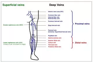
Sonographic Assessment of the Inferior Vena Cava. Alexis P Langsfeld MD. Anatomy . Anatomy . The inferior vena cava returns blood from the body to the right atrium Formed by the convergence of the illiac veins Retroperitoneal Right of the aorta Normal size <2.5 cm Varies w respiration.

E N D
Sonographic Assessment of the Inferior Vena Cava Alexis P Langsfeld MD
Anatomy • The inferior vena cava returns blood from the body to the right atrium • Formed by the convergence of the illiac veins • Retroperitoneal • Right of the aorta • Normal size <2.5 cm • Varies w respiration
Respiratory variation • expands w/ expiration • Contracts w/ inspiration • Due to changing intrathoracic pressures.
Probe Selection • Low frequency 2-5 MHz • Curvalinear probe
Orientation • Measure at the subcostal window • Adjacent to the rt atrium • Measure just below the diaphram - about 1 cm before the hepatic viens. • Parallel walls
Views • Cross section • Longitudinal
Aorta Thick echogenic walls Pulsatile High flow velocity Not compressable No respiratory variation IVC Thin walls Not pulsitile Low flow velocity Compressable Respiratory variation Aorta vs IVC
Flow Velocities • Aorta = High flow velocities • IVC = lower flow velocities
Benefits • Assessment of volume status • Find clots • Non-invasive • No radiation • At the bedside
Gestalt method • IVC Collapse with inspiration • “YES / NO” • Yes - CVP approximately 8 • No - CVP around 15
Research: ED Physicians Capable • Hruda, Jarslav et al. Echocardiographic Assessment of Preload Conditions Does Not Help at the Neonatal Intensive Care Unit. American Journal of Perinatology 2003;20:297-303. • Lloyd, TR. Accuracy of Central Venous Pressure Measurement From the Abdominal Inferior Vena Cava. Pediatrics 1992; 89: 506-508 • Krause, Irit et al. Inferior vena cava diameter: a useful method for estimation of fluid status in children on haemodialysis. Nephrology Dialysis Transplantation 2001; 16:1203-1206. • Lyon, Matthew, MD. Sonographic measurement of the inferior vena cava as a marker of blood loss. The American Journal of Emergency Medicine 2005; 23:45-50.
Research: CVP Assessment • Chen, Lei, Kim, Yunie, Santucci, Karen A. Use of Ultrasound Measurement of the Inferior Vena Cava Diameter as an Objective Tool in the Assessment of Children with Clinical Dehydration. Acad Emerg Med 2007 14: 841-845 • Krause, Irit et al. Inferior vena cava diameter: a useful method for estimation of fluid status in children on haemodialysis. Nephrology Dialysis Transplantation 2001; 16:1203-1206. • Lyon, Matthew, MD. Sonographic measurement of the inferior vena cava as a marker of blood loss. The American Journal of Emergency Medicine 2005; 23:45-50.
Conclusion • Anatomy review • Physiologic review • Technical review for good IVC measurements • Data supports use of IVC sonography as a rudimentary assessment of volume status • Can use clinically to guild patient management
References • Hruda, Jarslav et al. Echocardiographic Assessment of Preload Conditions Does Not Help at the Neonatal Intensive Care Unit. American Journal of Perinatology 2003;20:297-303. • Lloyd, TR. Accuracy of Central Venous Pressure Measurement From the Abdominal Inferior Vena Cava. Pediatrics 1992; 89: 506-508 • Krause, Irit et al. Inferior vena cava diameter: a useful method for estimation of fluid status in children on haemodialysis. Nephrology Dialysis Transplantation 2001; 16:1203-1206. • Lyon, Matthew, MD. Sonographic measurement of the inferior vena cava as a marker of blood loss. The American Journal of Emergency Medicine 2005; 23:45-50. • Textbook of Cardiovascular Medicine, Topol, Califf, Lippincott Williams& Wilkins 2006 • Color Duplex Sonography: Principles and Clinical Applications, Karl-Jürgen Wolf, Franz Fobbe, 2005, p.180-195 • Differential Diagnosis in Ultrasound, Gunter Schmidt, Pri, Werner, Asim Kurjak, Barbara (CON) Beuscher-Willems, - Medical - 2006