Download
1 / 74
840 likes | 1.51k Views
Pneumothorax. Mark Miller et al. Outline. Classification of Pneumothoraces Epidemiology & Pathophysiology Diagnosis Guidelines & Evidence Management options Summary. Case. 21 year old man presents to ED with left sided respirophasic chest pain. Differential diagnoses?. Case.

E N D
Pneumothorax Mark Miller et al.
Outline • Classification of Pneumothoraces • Epidemiology & Pathophysiology • Diagnosis • Guidelines & Evidence • Management options • Summary
Case • 21 year old man presents to ED with left sided respirophasic chest pain. • Differential diagnoses?
Case • 21 year old man presents to ED with left sided respirophasic chest pain. • Differential diagnoses? • Chest wall • Pleura • Lung • Pericardium • Mediastinum • Neuropathic
Case • Other important history?
Case • Other important history? • Physical Examination?
Case • Other important history? • Physical Examination? • Investigations?
Case • What’s the plan?
Case • What’s the plan? • Do an invasive procedure? • Do “nothing”?
Classification of Pneumothorax Spontaneous; • Primary spontaneous PMTX - occurs in people without clinically apparent lung disease (subpleural blebs & bullae) • Secondary spontaneous PMTX- occurring as a complication of preexisting lung disease (esp COAD)
Classification of Pneumothorax Other; • Iatrogenic (procedural- lines & biopsies) • Traumatic (Penetrating, blunt, barotrauma) • Non-Pulmonary in origin (Boerhaave’s, Catamenial, 2o to pneumoperitoneum)
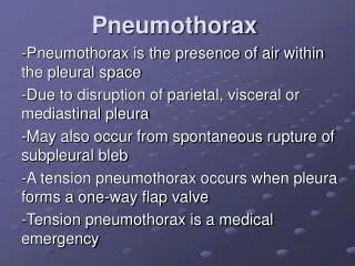
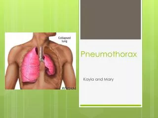
Pathophysiology • Lung tissue breach, air enters pleural space (negative pressure), intra-pleural pressure equilibrates with intra-bronchial pressure. • Unventilated segments create a shunt and potentially hypoxia. • Tension PMTX is due to “Flap-valve” effect, mediastinal shift distorts great vessels, reduces VR and causes hypotension.
Symptoms and signs. • Primary; • often occur at rest with ispilateral pain or acute SOB • symptoms may resolve within 24 hrs • small PTX (<15%) may have no signs, large PTX classic signs. Tachycardia is common. • ABG may show ⇑ A-a gradient and acute respiratory alkalosis
Symptoms and signs. • Secondary; • Small PMTX often badly tolerated in severe CAL- potentially life-threatening. • SOB and hypoxia often disproportionate to size of PMTX. Most have ipsilateral Pain • Signs often subtle & masked by underlying disease • hypercarbia often occurs
Symptoms and signs. • Signs include: • Asymmetrically decreased breath sounds • Hyperresonance • Altered vocal resonance • Often chest examination is unremarkable • Less commonly: Hamman’s crunch or Surgical emphysema • Tracheal deviation & shock= ?Tension pneumothorax
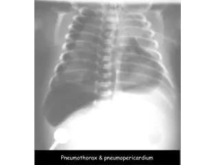
Complications. Acute; • Tension pneumothorax • Sc emphysema • Pneumomediastinum/ pericardium Delayed; • Failure to rexpand • Recurrence
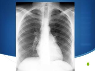
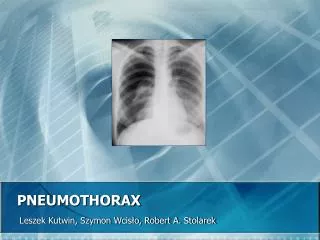
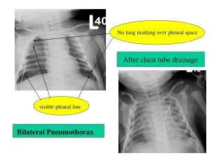
Diagnosis CXR • PA erect CXR will demostrate 83% • routine use of expiratory films doesn’t increase yield (?useful for small apical PTX). • Hyperlucency, lack of peripheral markings, fine visceral pleural line paralleling chest wall, blunted CP angle • lat. decubitus view may be helpful • supine films, PMTX easily missed - deep sulcus (CP angle)
Diagnosis • Estimating size of PTX is challenging and unecessary; • Small= <2cm between lung and chest wall • Large= >2cm between lung and chest wall
Treatment Options • Depends on pneumothorax size, symptom severity, co-morbidities, resources and patient preferences: Observation (& Oxygen) Aspiration Intercostal tube drainage Referral to specialists (physicians/surgeons)
Guidelines. • BTS Thorax 2003;58(Supp II):ii39-ii52 • ACCP Chest 2001; 119:590-602
Figure 1 Recommended algorithm for the treatment of primary pneumothorax.
Figure 2 Recommended algorithm for the treatment of secondary pneumothorax.
Evidence? • This is a data-poor area • Cochrane RV; Simple aspiration vs ICC for PSP 2007. • Based on 6 studies only, numbers are small, one RCT only (n=60)!!! • There is no significant difference in outcome success • Aspiration may reduce hospitalisation.
Evidence? • BMJ Evidence Review; • Small tubes are easier to insert but large PMTX may not respond. • Suction? Studies are too small and underpowered to show any benefit. • Optimal timing of pleurodesis is unclear.
Observation. • Sensible pts living close with small (<2cm) PSP who have minimal symptoms may be discharged with early outpatient review (?re X-Ray in ED at 24 hrs and 7days). • These patients should be advised that they should return in the event of worsening breathlessness or pain (no flying or diving). • up to 40% require further drainage
Denitrogenation... • Whilst in hospital (ED or ward) give high flow oxygen (10 L/min, not for COAD). • Resorption is 2% per day on room air – supplemental oxygen increases this rate 4-fold (Northfield TC BMJ 1971 –n=22) in humans plus several animal models.
Simple Aspiration • Big advantages… • Easier, less painful, involves fewer complications and may allow discharge home (fewer hospital days) • NB; there are small catheters that can be used for aspiration followed by UWSD if necessary.
Simple Aspiration • First line treatment for all primary pneumothoraces requiring intervention (59-83% success rate). • Only recommended as an initial treatment in small (<2cm) secondary pneumothoraces in minimally breathless patients under the age of 50 years (or as a temporary manoeuvre in the anti-coagulated).
Simple Aspiration • Use a seldinger kit (custom pigtail or CVL) and a 60mL leur lock syringe with a 3-way tap (avoid cannulas). • Aspirate until discomfort is felt, patient coughs, no more air aspirated , or 3L aspirated (leave catheter in situ) • Re-Xray now and at 6 hours
Simple Aspiration • What is success? Reduction to < 20% maintained at 6 (?) hrs. • If successful, re-Xray at 6 hours then discharge to follow up (back in 24hrs for X-ray then 7 days) • Admit successfully aspirated SSP
Simple Aspiration • Immediate failure - no/minor improvement. (if >2.5L aspirated then failure likely) • ? Role of repeated attempt (up to 20% success quoted)
Simple Aspiration In whom is aspiration less likely to be sucessful? • Large PMTX- some studies report correlation with size (82% success with moderate vs 43% with large- Aplin Emerg Med 1996) • Secondary PMTX • ? Earlier presentation • Age >50yrs
Simple Aspiration Pleurocath CVL Cannula
Simple Aspiration 14Fr Seldinger Pigtail catheter
Intercostal catheter drainage When? • If simple aspiration fails in PSP. • Secondary pneumothorax (except small PMTX in <50yrs). • Mechanical/NI ventilation planned. • Significant fluid/blood collection • Tension PMTX • Bilateral PMTX • Air transport planned • Recurrence if chemical pleurodesis planned
Intercostal catheter drainage What Size? • There is no evidence that large tubes (20-24F) are any better than small tubes (10-14F). • The initial use of large tubes is not recommended, although it may become necessary if there is a persistent air leak.
Intercostal catheter drainage Methods • A brutal, torturous procedure in the wrong hands. • Seldinger style tubes. • Blunt dissection- finger into pleural space NO TROCAR! • Use maximum volume of LA allowable and infiltrate en route through the chest wall. • Use morphine and sedation if possible • In most adults a depth of >12cm at skin is required.