Download
1 / 18
180 likes | 309 Views
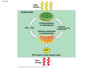
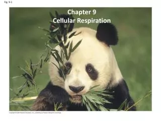
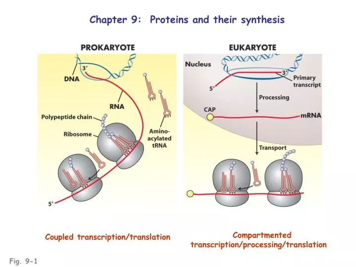
Chapter 9: Proteins and their synthesis. Compartmented transcription/processing/translation . Coupled transcription/translation . Fig. 9-1. Proteins are polymers of amino acids joined through peptide bonds. Each protein has a amino- and a carboxyl-terminus. Fig. 9-2.

E N D
Chapter 9: Proteins and their synthesis Compartmented transcription/processing/translation Coupled transcription/translation Fig. 9-1
Proteins are polymers of amino acids joined through peptide bonds Each protein has a amino- and a carboxyl-terminus Fig. 9-2
The function of a protein is dependent upon its overall structure; each level of protein structure is dependent upon lower levels; thus, all are derivatives of primary structure of the polypeptide The primary structures of polypeptides are directly derived from theprimary structures of their mRNAs Primary structures of mRNA are derived from the primary structures of their DNA templates(± splicing) Changes in DNA sequence can alter function of proteins encoded by that DNA sequence
Protein higher order structures amino acid sequence Regular coil/sheet motifs stabilized by H-bonds between peptidyl atoms specific intramolecular folding stabilized by associations of amino acid “R” groups intermolecular associations stabilized by associations of amino acid “R” groups Tertiary and quaternary structures are determined by primary structure Fig. 9-3
Genes encode the primary structure of proteins E. coli trpA mutations: genetic map is co-linear with the protein (C. Yanofsky) Fig. 9-5
Degeneracy in the codon-amino acid code derives from: • Multiple codons for certain same amino acids • e.g., UCU • UCC • UCA serine • UCG • UGU • UGC
tRNAs convert the codon-amino acid code mediated by aminoacyl synthetases Fig. 9-10
Degeneracy in the codon-amino acid code derives from: • Multiple codons for certain same amino acids • e.g., UCU • UCC • UCA serine • UCG • UGU • UGC • “Wobble” permits certain individual tRNAs to pair with • multiple codons
“Wobble” creates partial ambiguity in codon 3’ nucleotides Fig. 9-12
Three significant domains of ribosome during translation: A: incoming aminoacyl-tRNA binding site P: peptidyl-tRNA binding site E: exiting deacylated-tRNA site Fig. 9-15
Three phases of translation: • Initiation: • association of small subunit and • capped 5’ end of mRNA • association of Met-tRNA (fMet) • scanning to AUG (eukaryotes) • association of large subunit Fig. 9-18
Three phases of translation: • Initiation • Elongation • aa-tRNA association • with A site • transfer of peptidyl • to aa-tRNA • translocation (next codon) • exiting of deacylated tRNA Fig. 9-19
Three phases of translation: • Initiation • Elongation • Termination • stop codon recruits • release factor • hydrolysis of peptidyl-tRNA link • release of complex Fig. 9-21
Translational suppressors: mutant tRNAs with modified anticodons that permit “readthrough” of nonsense mutations Fig. 9-23
Posttranslational modifications of proteins: • protein folding into “native” configuration (assisted by chaperones) • covalent modifications of amino acid side chains • targetting to specific intra- and extracellular sites • All subject to mutation in the protein or in the cellular machinery that modifies the protein