Download
1 / 25
250 likes | 563 Views
Status Epilepticus. PICU Resident Lecture Series Lucile Packard Children’s Hospital (Updated: April 2011). Objectives. What are common causes of SE Learn the physiologic sequela of SE (Why do these patients need to be in the PICU?) Learn what tests/labs are needed acutely

E N D
Status Epilepticus PICU Resident Lecture Series Lucile Packard Children’s Hospital (Updated: April 2011)
Objectives • What are common causes of SE • Learn the physiologic sequela of SE • (Why do these patients need to be in the PICU?) • Learn what tests/labs are needed acutely • Acute management of SE • Including procedures, medications, and “pentobarb” comas
Definitions • No absolute definition of Status Epilepticus (EP) • Generally accepted definition is • Greater than 30 minutes OR • Frequent seizures without returning to baseline • Treatment if seizure lasts >5 minutes • High risk of lasting >30 minutes • Delayed treatment can lead to permanent sequela
Common drugs related to seizures • Penicillins • Isoniazid • Metronidazole • Antihistamines • Narcotics • Ketamine • Halothane/Enflurane • Tricyclic antidepressants • Antipsychotics • Phencyclidine • Cocaine
Physiologic Consequences of SE • Phases of SE • Respiratory Effects • Hyperpyrexia • Metabolic derangements • Laboratory changes • Summary
Phases of SE • Hyperdynamic Phase • Increased cerebral metabolic demand • Massive catecholamine/autonomic discharge • Increased CBF, HTN, tachycardia • Exhaustive Phase (with persistent SE) • Catecholamine depletion • Hypotension, decreased CBF • Can lead to neuronal damage (ongoing metabolic demand with tissue hypoxia)
Respiratory Effects • Hypoxia and Hypercarbia are common • Chest wall rigidity (muscle spasms, oral secretions) • Hypermetabolic state with increased 02 demand and increased C02 production • Neurogenic pulmonary edema is rare complication • Marked increased in pulmonary vascular pressure is presumed etiology
Hyperpyrexia • Can lead to seizures or be a result of SE • Exacerbates mismatch of cerebral metabolic demand and substrate delivery • Therefore fevers should be treated aggressively • Antipyretics/cooling
Metabolic derangements • Acidosis • Lactic acidosis due to poor tissues oxygenation with inc energy expenditure • Respiratory acidosis may also develop • Glucose • Initial hyperglycemia from catecholamine surge followed by hypoglycemia • Can be detrimental to the brain, and can further worsen lactic acidosis
Metabolic derangements (cont’d) • Rhabdomyolysis • Protracted tonic-clonic activity can have extensive muscle breakbdown • Leads to hyperkalemia, myoglobinuria • Leukocytosis • Stress response causes demarginalization of SBCs • In 15% of children, this leukocytosis can be seen in the CSF
Treatment • ABCs • Venous access • Labs • Other diagnostic • Meds
ABCs • Avoid hypoxia by providing oxygen (facemask or NC) • Oral airway can be helpful (but difficult to place) • Nasal trumpet is good alternative • Optimize position, jaw thrust • If poor respiratory effort, begin bag-mask ventilation and consider intubation
Intubation • Some indications: • Difficult to maintain airway • Unable to manage oral secretions • Ineffective respiration • Hypoxia • Hypercarbia • CNS pathology, unequal pupils • SE >30 minutes despite appropriate treatments • REMEMBER: paralytics DO NOT control CNS epileptiform discharges
Venous access • Obtain IV/IO access • Can give IM or Rectal meds but venous access is necessary • Blood pressure management • Hypertension likely to resolve with sz control • Some cases need tx (like inc BP with renal failure) • Start volume resuscitation if hypotensive with bolus of NS (20ml/kg)
Labs required in ALL pts with SE: • CBC, Chem panel (with LFTs, glucose, ca, mg) • Hyponatremia and hypocalcemia are readily treatable • Stat beddside glucose (*especially in neonates and infants) • Ammonia • Anticonvulsant levels • Tox screen • LP: defer in pts with signs of increased ICP or if unstable (but do not delay therapy i.e. abx)
Other diagnostics • CT scan • Focal seizures or deficits; History of trauma • Non-contrast: mass lesions, hemorrhage, hydrocephalus • Contrast: meningitis, abscess, encephalitis • EEG- indicated in ALL pts with SE • Standard: one time study in SE that has resolved • Continuous: difficult to control SE, burst suppresion, subclinical seizures • Video: can be used in conjunction for seizures that are difficult to characterize
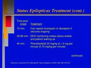
Medications • Initiate antiepileptic therapy early • With delayed treatment, pt will also have delayed response to treatment • Thus requiring higher doses • Combine rapid acting to control with long acting to prevent recurrence
Persistent SE • “Pentobarb” coma • CNS electrical quiescence by continuous infusion • Pentobarbital: 1-3mg/kg/hr after bolus (10mg/kg) • Midazolam: 1-10mcg/kg/min after bolus (0.15mg/kg) • Propofol 20-70 mcg/kg/min • Normal physiologic activity also suppressed • Intubation necessary
“Pentobarb” coma (cont’d) • Central line placement • For delivery of continuous infusion • May cause hypotension so pt may require rapid fluid bolus or inotropes • Treat hypotension aggressively in these pts • Continuous EEG • “Burst suppression” is the specific electric pattern noted on EEG once in a successful coma. Electrical activity is only noted once per screen (15-20sec)
“Pentobarb” coma (cont’d) • Pt must be started on a long acting anticonvulsant • Check for therapeutic levels • Burst suppression for 24-48 hrs • Coma gradually lifted while monitoring for seizure activity
Non-convulsive SE • Up to 20% of children with SE have non-convulsive SE after tonic-clonic activity • If no response to painful stimulation within 20-30 min of tonic-clonic activity • Urgent EEG • Must maintain High Index of Suspicion • Often difficult to assess (i.e. previous medications, post-ictal state) • Neurology consult is imperative