Download
1 / 67
690 likes | 1.07k Views
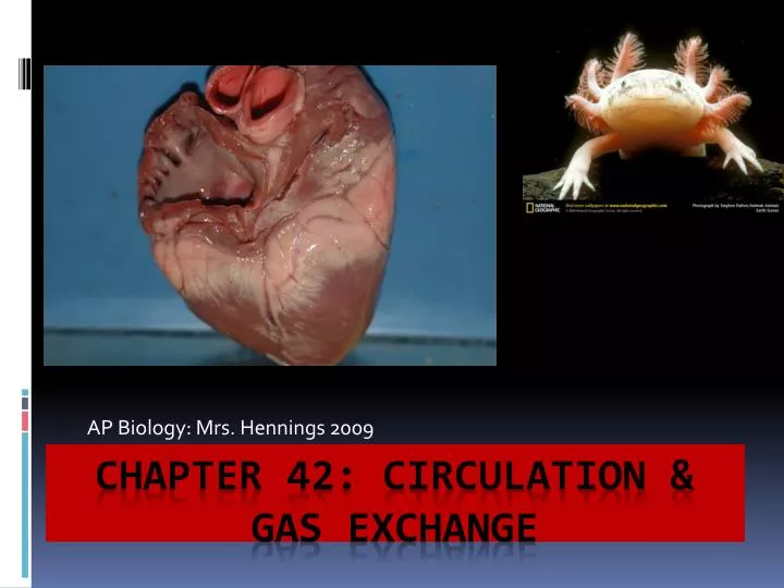
AP Biology: Mrs. Hennings 2009. Chapter 42: Circulation & gas exchange. Trading Places…. All animals need to exchange substances with their environments This exchange ultimately happens at the cellular level In multicellular animals there are specialized systems to carry out exchange.

E N D
AP Biology: Mrs. Hennings 2009 Chapter 42: Circulation & gas exchange
Trading Places…. • All animals need to exchange substances with their environments • This exchange ultimately happens at the cellular level • In multicellular animals there are specialized systems to carry out exchange
Remember Diffusion??? • Small nonpolar molecules such as oxygen and carbon dioxide can move between cells and their surroundings by diffusion • Diffusion is a very slow process for more than a few mm distance • Natural selection – 2 adaptations • Body size keeps most cells in contact with environment • Circulatory system
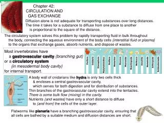
Gastrovascular cavities • Some animals don’t have a circulatory system • Hydras and other cnidarians • Planarians and other flatworms
Open & Closed Circulation • Animals with lots of cell layers- too thick for diffusion • Circulatory system minimizes distance of exchange • A circulatory system has 3 parts: • Circulatory fluid • Interconnecting tubes • Muscular pump- the heart
Open Circulatory System • Arthropods and mollusks • Circulatory fluid baths organs directly • Fluid is called “hemolymph”
Closed Circulatory System • Blood confined to vessels and is separate from interstitial fluid • High blood pressures- fast delivery
Evolution of vertebrate circulatory system fish amphibian reptiles birds & mammals 2 chamber 3 chamber 3 chamber 4 chamber Birds ANDmammals! Wassssup?! V A A A A A A A V V V V V
Cardiovascular System • Closed circulatory system • Arteries, veins, capillaries • Arteries- blood away from heart to organs • Arterioles- in organs smaller branches of arteries- carry it to capillaries. • Capillaries- microscopic with capillary beds • Venules- and Veins- carry blood back to heart
Which way to travel??? • Arteries – carry blood AWAY from the heart.Veins – carry blood TO the heart Hearts of all vertebrates have atria (receive) And ventricles that pump out
Single circulation • Bony fish, ray, sharks • Two chambers- an atrium and ventricle
Double circulation • Amphibians • Reptiles • Mammals • Pumps for 2 circuits serve different tissues but combined into 1 heart
Adaptations of Double Circulatory Systems • Amphibians: Heart with 3 chambers: • Two atria and one ventricle • Ridge in ventricle diverts 90% of oxygenated poor blood from RA to the pulmonary circuit- and most of the oxgen rich blood from LA to the systemic circuit • When underwater- a frog shuts off lung circulation- and blood flow continues under the skin.
Amphibian Circulation Summary • Three chambered heart • 2 atria and one ventricle • Two circuits of blood • Pulmocutaneous • Systemic
Reptiles ( Except for birds) • Lizards, snakes, turtles- • 3 chambered heart • Septum partially divides single ventricle • In crocodilians the septum is complete so they have 4 chambers
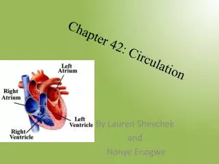
Mammals and birds • Ventricle is completely divided • 2 atria and 2 ventricles • Left side receives and pumps oxygen rich blood • Right side receives and pumps oxygen poor blood • Powerful 4 chambered heart supports endothermic life
Mammalian Circulation • Right ventricle- contracts- blood goes to lungs via the pulmonary arteries • Blood flows through capillaries in lungs and loads up with oxygen and unloads carbon dioxide • Oxygen rich blood goes back to heart via the pulmonary veins • It is dumped into the left atrium
Mammalian circulation Contd. • It flows from LA to LV and then is pumped out to the body tissues • Blood goes out of the LV via the aorta • The first branches off the aorta are the coronary arteries ( the best for the heart) • Branches lead to head and arms (ascending) • Branches lead to abdomen (descending) and legs • Oxygen poor blood in head and neck- superior vena cava into RA • Oxygen poor blood in legs etc- inferior vena cava into RA
Coronary arteries bypass surgery
How does the heart work? • Behind sternum- breastbone • Size of clenched fist • Mostly made of cardiac muscle • Two atria • Two ventricles • Contracts and relaxes in rhythmic cycle • One complete sequence of pumping and filling is the cardiac cycle
The contraction phase is called systole • The relaxation phase is called diastole • The volume of blood pumped per min is called the cardiac output- normal 5 L/min • There are two things that determine cardiac output • Heart rate ( per min) • Stroke volume ( amount of blood pumped by a ventricle in one contraction) – 70 mL
There are valves- keep blood moving in correct direction • Made of flaps of connective tissue • AV valve- between each atria and ventricle • Semilunar valves- at 2 exits of heart where aorta leaves ventricle and pulmonary artery leaves right ventricle • Heart sounds- Lub= blood against closed AV valves • Dub= recoil of blood against semilunar • Heart murmur- if blood squirts back by defective valve
Cardiac Output Amount of blood pumped by each ventricle in one minute CO = Heart rate X Stroke volume Stroke volume—amount of blood ejected from ventricle with each heart beat—70 ml 75 bpm X 70 ml = 5250 ml/min (5.25L)
Heart murmurs • Some congenital • Damaged by rheumatic fever • Group A strep bacteria • Endocarditis • Can replace valve if needed
Keeping the beat… • Heartbeat starts in heart • Cardiac cells are autorhythimic without signal from nervous system • Heart cells removed from heart beat in a dish! • Group of cells in RA- SA node- pacemaker • Sets rate and timing • SA node generates electrical signal • EKG can measure currents
SA node impulse spreads through atria- contract together • AV node- slight delay before reach ventricles • Delay allows atria to empty • AV node then conducts to ventricles where special muscle fibers called bundle branches and Purkinjie fibers conduct
Control of the beat… • Physiological cues- regulate SA node • Two sets of nerves: sympathetic and parasympathetic • One set speeds up pacemaker and other slows it down • Stand up- sympathetic nerves increase HR • Sit down- parasympathetic nerves decrease HR • Hormones influence- EPINEPHRINE • Body temperature faster if fever
Blood Flow • Basic Blood Vessel Structure & Function • Central lumen ( cavity) lined with endothelium- minimizes resistance to blood flow • Surrounding endothelium specialized tissues that differ depending on job of vessel
Blood vessels arteries veins artery arterioles venules arterioles capillaries venules veins
capillaries • Smallest blood vessel • Diameter only slightly larger than RBC • Thin walls • Endothelium and basal lamina • Structure facilitates exchange of substances
Arteries • More complex than capillaries • Two layers of tissue surrounding endothelium • Outer layer of connective tissue- elastic • Middle layer- smooth muscle • Wall is 3 times as thick vs. vein • Can pump at high pressure • Signals from nervous system and hormones
Veins • 2 layers of tissue-like arteries • Thinner walls compared to arteries • Valves maintain one way blood flow
Velocity of Blood • What influences flow rate? • Diameter of hose! • Blood slows from arteries to capillaries- because there are many more capillaries compared to large vessels. • Blood moves 500 times slower in capillaries compared to aorta!
Blood Pressure • Moves from high pressure to low pressure • Contractions of heart make pressure • Arterial pressure highest when heart contracts called systolic pressure • Pulse- heart rate- • When ventricles relax- diastolic pressure
Regulation of BP • Two time scales- • Cardiac cycle oscillations • Longer time scale • Vasoconstriction- smooth muscles in arteriole walls contract- increases BP • Vasodilatation- increase in diameter BP decreases
Measurement of blood pressure • High Blood Pressure (hypertension) • if top number (systolic pumping) > 150 • if bottom number (diastolic filling) > 80
Fluid Return by Lymph System • 85% of fluid that leaves capillaries is returned to capillaries • Imbalance loss of 4L of fluid from capillaries to tissues • Lost fluid and proteins return to blood system via the lymphatic system • Lymph- drains into circulatory system • Lymph Nodes- organs- filtering lymph and house to cells that are fighters
Lymphatic system • Parallel circulatory system • transports white blood cells • defending against infection • collects interstitial fluid & returns to blood • maintains volume & protein concentration of blood • drains into circulatory system near junction of vena cava & right atrium